Download
1 / 15
150 likes | 247 Views
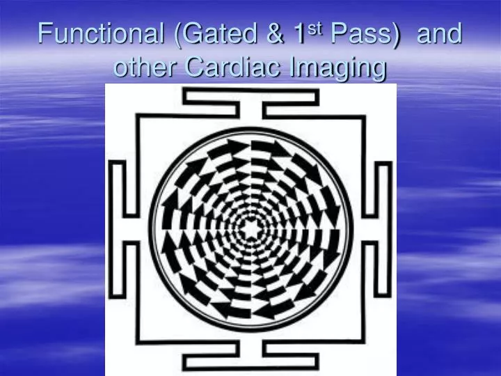
Functional (Gated & 1 st Pass) and other Cardiac Imaging. Coronary Artery Perfusion. Left main coronary artery Left anterior descending artery I. V. septum L. V. anterior wall Left circumflex Left atrium L. V. posterior L. V. lateral wall Right coronary artery Right atrium

E N D
Coronary Artery Perfusion • Left main coronary artery • Left anterior descending artery • I. V. septum • L. V. anterior wall • Left circumflex • Left atrium • L. V. posterior • L. V. lateral wall • Right coronary artery • Right atrium • Right ventricle • L. V. inferior wall
Mechanical Activity • Systole – ventricular contraction • Diastole – ventricular relaxation
1st pass protocol Patient Preparation • NPO 4-12 hours • No caffeine Procedure • Anterior or 45 LAO • Gated or non-gated • LFOV camera • LEHR collimator • 20 mCi pertechnetate, DTPA, Sestamibi • Good bolus ( <1.0 ml) – follow with saline flush • List mode (16 frames/cycle) or Frame mode (20-35 frames/sec) • 64x64 matrix • 1-2 minutes total acquisition time.
MUGA (Multiple Gated Acquisition) Provides functional information Patient preparation • NPO 4-12 hours • No caffeine Blood preparation • In-vivo • Inject cold PYP followed ~20 minutes later by 20-30 mCi Tc-99m • In-vivtro (modified In-vivo) • Inject cold PYP; withdraw 3-5 mLanticoagulated blood into 10 mL syringe containing ~25 mCi Tc-99m; incubate 10 minutes then re-inject. • In-vitro commercial kits (UltraTag) RBC)
MUGA Imaging Procedure • ECG • Anterior and ~45 LAO (best septal separation) w. caudal tilt • 8 to 16 frames/second • 64x64 matrix • 10 minutes/view
Processing • EF (ejection fraction) = (ED-ES)/ED x 100 ED = end-diastolic volume ES = end-systolic volume • SV (stroke volume) = ED-ES • CO (cardiac output) = SV x heart rate
Myocardial Infarct Imaging15-30 mCiTc-99m Pyrophosphate • Of historical interest – replaced by serum enzyme testing
Positron Emission Tomography • Myocardial perfusion • Rubidium-82 chloride • Nitrogen-13 ammonia • Oxygen-15 water • Myocardial metabolism • Fluoride-18 FDG • Carbon-11 palmitate • Carbon-11 acetate