Equine Lameness
Equine Lameness . Equine Lameness Exam. One of the most common (if not the #1) body systems evaluated and treated is the musculoskeletal system Detecting the source of lameness can be daunting – many probs. have no obvious external signs Common Clinical Signs include swelling, heat

Equine Lameness
E N D
Presentation Transcript
Equine Lameness Exam • One of the most common (if not the #1) body systems evaluated and treated is the musculoskeletal system • Detecting the source of lameness can be daunting – many probs. have no obvious external signs • Common Clinical Signs include • swelling, heat • Discharge • muscle atrophy • lameness (#1)
Equine Lameness Exam • 3 reasons for lameness include: • Pain (#1) • Mechanical interference w/out pain (scar tissue) • Neurological
Equine Lameness Exam • 3 goals of a lameness exam • Identify the location • Diagnose • Treatment plan
Equine Lameness Exam • 1ststep is to obtain a complete history - Signalment - Length of issue - Previous health issues - Speed of onset - Exercise induced - Known trauma - Any treatment started - Pattern to the lameness
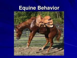
Equine Lameness Exam • Next the horse is observed at rest & in motion • Rest • Observe from a distance for any obvious abnormalities, confirmation, how horse stands (holds legs) • Motion • Observe horse walking to & from the clinician, may need multiple surface types, may need to remove shoes, observe head & neck carriage
Equine Lameness Exam • Motion (cont.) 1. Walk • In a straight line • Up & down an incline • Backing up 2. Trot – usually the most informative gait • In a straight line • In a circle (both directions) • Flexion tests
Equine Lameness Exam • Palpation – feeling for any heat, swelling, or pain • The wear pattern of the hoof or shoe is evaluated • Hoof test for pain • Nerve blocks may be used to localize the area of pain
Equine Lameness Exam • Misc. tests include - X-rays - Ultrasound - Thermography - Nuclear scintigraphy - MRI - CT - Arthrocentesis - Rectal exam - Biopsy - Force plate gait analysis - High speed cinematographic gait analysis
Equine Lameness • Predisposing factors to lameness • Heredity– Very few are directly inherited, but confirmation types that often lead to lameness are inherited (small feet, straight pasterns, cow-hocked) • Congenital– Bone, tendon, joint, & ligament development may be impaired while in utero
Equine Lameness • Predisposing factors to lameness cont’d: • Negligent or improper foot care • infrequent trimming • unbalanced trimming • poor fitting shoes • shoeing aids
Equine Lameness • Predisposing factors to lameness cont’d: • Improper training methods or over training • over use of a lunge line • lunging in small circles • one direction lunging • poor footing in training area • training too early, training too rapidly
Equine Lameness • Predisposing factors to lameness • Nutrition of the growing horse – feeding high levels of protein, improper mineral content, overweight • Wounds • Overuse – racing, jumping, barrel racing, roping
Equine Lameness http://www.youtube.com/watch?v=zH4YySG1D_w http://www.youtube.com/watch?v=n4B8yNJUn-U
Equine Lameness • The Laminae is a structure between the hoof wall and coffin bone (P3) composed of a network of interlocking blood vessels and tissue (epidermis) that serve to connect the hoof to the foot and to provide blood supply
Laminitis/Founder • Equine laminitis is a vascular disease • Associated with areas of ischemia or hemostasis within the laminae • The laminae secure the coffin bone/distal phalanx to the hoof wall
Laminitis/Founder • Inflammation associated with delamination interferes with the wall/bone bond • In advanced laminitis, the coffin bone becomes detached from the horny wall and may rotate or sink. • In lay terms, this is known as “founder”
Laminitis • Three phases of laminitis in horses are identifiable: • Developmental • Acute • Chronic
Laminitis • Since pre-existing illness leads to laminitis, the symptoms of early laminitis are also the symptoms of the precipitating illness. • Digital pulses and distal limb temperatures may be increased or decreased but no lameness is evident • Occasionally, no development phase can be recognized; the horse is simply found to be in the acute phase with no apparent ill health preceding or accompanying it
Laminitis - Treatment • Treatments for laminitis vary according to the severity of the condition but include: • Encouraging the horse to lie down to relieve pressure on the hoof/hooves. • Imposing dietary restrictions to prevent overeating and obesity. • There is a strong link between excess blood Glucose and laminitis • Administering fluids if the horse is ill or dehydrated. • Administration of painkillers, since moderate to intense pain often accompanies laminitis and founder