Download
1 / 14
140 likes | 169 Views
Comprehensive study on methylation & gene expression in cervical cancer cells post-treatment, with visualization & statistical analysis.

E N D
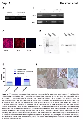
Sup. 1 Huisman et al A B C D E F untreated 5-aza-dC treated m u m u untreated 5-aza-dC treated m u m u HeLa SiHa CaSki CaSki C33A HeLa C33A HeLa 42 kDa 42 kDa HeLa CaSki C33A Maspin Actin * Figure S1 (a)Maspin promotor methylation status before and after treatment with 5-aza-dC (5 µM) in C33A cells visualized with a MSP. (b) C13ORF18 promotor methylation status before and after treatment with 5-aza-dC (5 µM) in C13ORF18-methylated HeLa and SiHa cells visualized with a MSP. For MSP, PCR primers specific for methylated template (m) and unmethyalted template (u) are shown. Maspin expression in cervivcal cancer cells as analyzed with ICC (c) and western blot (plus Actin loading control) (d) in HeLa, CaSki and C33A. (e)Quantification of the methylation status of the Maspin promoter in DNA obtained from cell lines, normal cervices and cervical cancer patients measured by MSP. Differences in methylation levels were quantified using ImageJ and the ration of the methylated to unmethyhlated primer pairs for each sample was determined. Samples were classified as being methylated (ratio of >1.1), intermediately methylated (ratio of 0.9-1.1) or unmethylated (ratio of <0.9). Statistical significance was determined with a MannWhitney U test and a value of 0.05 or less was considered statistical significant (* p<0.05). (f) Maspin IHC staining for four primary tumors (up) and metastatic tumors from four patients (down). The tumor-surrounding tissue served as control for base line levels of Maspin expression.
Sup. 2 Huisman et al TET1TET2 TET3 * * ** *** Figure S2 TET 1-3 expression in healthy control cells and cervical cancer cell lines. Endogenous expression of TET1, TET2 and TET3 in normal cells (healthy ovarian cells and fibroblasts) and cervical cancer cell lines. Values were quantified by qPCR and each bar represents the mean of in general three independent experiments measured in triplicate ± SEM. Statistical significance was determined using one-tailed t-test. A p-value of 0.05 or less was considered statistical significant (*p≤0.05, **p<0.01 and ***p<0.001).
Sup. 3 Huisman et al A B C ** ** *** ** ** *** * ** Figure S3 Endogenous re-expression of CCNA1, TFPI2 and Maspin mRNA by ATFs. (a) Relative expression of CCNA1 mRNA after treatment with CCNA1-targeting ATFs in the CCNA1-negative cell lines HeLa, SiHa, CaSki and C33A. (b) Relative expression of TFPI2 mRNA after treatment with TFPI2-targeting ATFs in the TFPI2-methylated cell lines HeLa and SiHa. (c) Relative expression of Maspin mRNA after treatment with Maspin-targeting ATFs in the Maspin-unmethylated cell lines SiHa and CaSki and the Maspin-methylated cell line C33A. Each bar represents the mean of in general three independent measured in triplicate ± SEM. Quantification of mRNA was performed using qRT-PCR and induction levels were normalized to GAPDH and relative to an empty vector. Statistical significance was determined using one-tailed t-test. A p-value of 0.05 or less was considered statistical significant (*p≤0.05, **p<0.01 and ***p<0.001).
Sup. 4 Huisman et al A B C D E F G CSCC-7 CSCC-8 CC-10 CC-11 50 µM 50 µM 50 µM 50 µM 10x 10x 10x 10x Figure S4 Visualization of the low passage cell lines CSCC-7, CSCC-8, CC-10 and CC-11. CSCC-7 and CC-11 grow as spherical colonies, while CSCC-8 and CC-10 grow as monolayer (a). Relative expression of C13ORF18 mRNA after treatment with C13ORF18-targeting ATFs (3ab-VP64 and 5ab-VP64) and aspecific ATFs in CSCC-7 (b), CSCC-8 (c), CC-10 (d) and CC-11 (E) cells. Relative expression of CCNA1 mRNA after treatment with the CCNA1-targeting ATF (12ab-VP64) and nonspecific ATFs in CSCC-7 (f) and CSCC-8 cells (g). Expression was normalized to GAPDH and relative to an empty vector. Bars represent the average ± SEM of a triplicate measurement.
Sup. 5 Huisman et al empty vector (background reads) 3ab targeting C13ORF18 3ab -10000 bp TSS +10000 bp -10000 bp TSS +10000 bp 12ab targeting CCNA1 43ab targeting TFPI2 12ab 43ab -10000 bp TSS +10000 bp -10000 bp TSS +10000 bp Figure S5 ZFP association with their aimed target site. Enrichment of C13ORF18- (3ab, (5ab next page)), CCNA1- (12ab) and TFPI2- (43ab) targeting ZFPs at their respective target sites in CaSki cells as detected by ChIP-Seq and visualized with RStudio. Also shown are the background reads at the target site after expressing an empty vector, and the off-target reads of the ZFPs at the promoters of the various other genes. ZFPs are aimed at the indicated target site ( ). Also shown is the TSS ( ).
Sup. 5 (continue) 5ab targeting 13ORF18 5ab -10000 bp TSS +10000 bp Figure legend on previous page.
Sup. 6 Huisman et al A B empty vector 3ab-NoEf 5ab-NoEf 12-NoEf 43ab-NoEf Chromosome1 2 3 4 5 6 7 8 9 10 11 12 13 14 15 16 17 18 19 20 212122 x y coverage ZFP target site HA-tag antibody background 12-NoEf empty vector 12-NoEf empty vector -10000 bp 1bp mismatch +10000 bp -10000 bp 1bp mismatch +10000 bp 12ab-NoEf target site CCC CGC CCA GCC GGC CAC 1 bp mismatch CCC CGC CCG GCC GGC CAC 12ab-NoEf target site CCC CGC CCA GCC GGC CAC 1 bp mismatch CCC CGC CCG GCC GGC CAC Figure S6 Genome-wide binding of the various gene-targeting constructs. (a) Binding patterns of 3ab, 5ab, 12ab, 43ab and background coverage (empty vector) to the whole genome as analyzed by ChIP-Seq. Global enrichment was visualized using NextGENEe software. (b) Coverage plots of two off-targets for 12ab-Noef, which correlate with a 1 bp mismatch (in red and underlined) in the intended target sequence of 12ab (shown below plots) on chromosome 14 and 20. Also shown is the coverage for the empty vector at this location.
Sup. 7 Huisman et al empty vector 12-NoEf chromosome 7: CCC CGC CCA GCC GGC CAC chromosome 11: CCC CGC CCA GCC GGC CGC chromosome 14: CCC CGC CCA GCC GGC CTG chromosome 20: CGC CGC CCA GCC GGC CAC -10000 bp mismatch +10000 bp -10000 bp mismatch +10000 bp Figure S7 Off-targets caused by sequence similarity. Coverage plots showing a local enrichment for 12ab-NoEf of 1 or 2 bp mismatches at the outer site of the target sequence of 12ab-NoEf (CCC CGC CCA GCC GGC CAC). Mismatched basepair(s) are indicated in red and underlined. Shown is a 20 kb spanning region and coverage is visualized with RStudio (cells expressing an empty vector were used as control (left)). Local enrichment was determined using the coverage distribution report obtained from NextGENe .
Sup. 8 Huisman et al regular transduction superinfected A B C D E regular transduction superinfected * * ** * Figure S8 Relative GFP mRNA expression after retroviral treatment with C13ORF18- (a) and CCNA1- (b) targeting constructs in CaSki. On the left, the GFP expression after regular transduction, on the right the GFP expression after superinfection. The targeting constructs carry either the VP64 activator domain, TET2-CD, TET2-mutant or NoEf. Relative C13ORF18 (c),CCNA1 (d) and TFPI2 (e) expression after retrovirally induced expression of the gene-targeting ZFPs carrying the transcriptional activator VP64 using the superinfection procedure. For superinfection, cells were sorted and re-transduced before analysis. Gene expression is relative to GAPDH. Each data point represents the mean ± SEM of three or more independent experiments measured in triplicate. Statistical significance was determined using a one-tailed t-test. A p-value of 0.05 or less was considered statistical significant (*p≤0.05 and **p<0.01).
Sup. 9 Huisman et al methylated CpG unmethylated CpG A B TSS 5ab-NoEf 5ab-TET2-CD 1 2 3 4 5 6 3ab-NoEf + 5ab-NoEf 3ab-TET2-CD + 5ab-TET2-CD 3ab-VP64+ 5ab-VP64 5ab-target site 3ab target site * ** ** Figure S9 Demethylation of the C13ORF18 promoter after ZFP-mediated targeting of TET2 or VP64. (a)DNA methylation status is shown for 39 CpGs in the C13ORF18 promoter after expressing 5ab-NoEf and 5ab-TET-CD or simultaneous expression of 3ab-NoEf/5ab-NoEf, 3ab-TET2/5ab-TET2-CD or 3ab-VP64/5ab-VP64 in CaSki cells. Each row represents an individual clone. Shown are the 3ab and 5ab target sites and the TSS. (b) The quantification of the DNA methylation of the 39 CpGs in the sequenced C13ORF18 promoter with the various constructs. Statistical significance was determined using a one-tailed t-test. A p-value of 0.05 or less was considered statistical significant (*p≤0.05 and **p<0.01).
Sup. 10 Huisman et al EPB41L3 5ab-NoEf A B -5 kb TSS +5 kb CpGs 1 2 3 4 5 6 7 8 9 10 11 12 13 14 15 12ab-NoEf EPB41L3 -5 kb TSS +5 kb ** CpGs 1 2 3 4 5 6 7 8 9 10 11 12 13 14 15 Figure S10. Off-target effects of TET-ZFPs on EPB41L3 promoter methylation. (a) The off-target effect of the C13ORF18-targeting 5ab-TET2-CD and the NoEf-control on the methylation status of the EPB41L3 promoter. (b) The off-target effect of the CCNA1-targeting 12ab-TET2-CD and the NoEf-control on the methylation status of the EPB41L3 promoter. Methylation levels were quantified by pyrosequencing and each data point represents the mean ± SEM of three independent experiments (same samples as from Figure 5). Inserts show the binding of the 5ab-NoEf and 12ab-NoEf to the EPB41L3 promoter as analyzed by ChIP-Seq and visualized by NextGene. Each gray block represents a coverage of one, and the blue line indicates the gene position. Statistical significance was determined using a one-tailed t-test. A p-value of 0.05 or less was considered statistical significant (**p<0.01).
Table SI Huisman et al Table SI Sequence of ZFPs targeting C13ORF18, CCNA1, TFPI2 and Maspin.Shown are the amino acid sequences of the six variabe regions of all ZFPs used in this study as obtained from zincfingertools.org. Below is the AA sequence of the ZFP backbone according to the Sp1 consensus framework plus the six variable regions (ZF1 till ZF6) in red (exemplified for 3ab). For cloning in the pMX-IRES-GFP, the protein coding regions were flanked with SfiI restriction sites (see M&M). 3ab AA sequence: LEPGEKPYACPECGKSFSTSGNLVRHQRTHTGEKPYKCPECGKSFSRSDKLTEHQRTHTGEKPYKCPECGKSFSRSDKLVRHQRTHTGEKPYACPECGKSFSQSGDLRRHQRTHTGEKPYKCPECGKSFSREDNLHTHQRTHTGEKPYKCPECGKSFSRSDKLVRHQRTHTGKKTS
Table SII Huisman et al Table II Sequences primers.
Table SII Huisman et al Table II Sequences primers (continuation of previous page).