Download
1 / 50
580 likes | 809 Views
Learn about the classification, common infections, pathology, diagnosis, and treatment of intestinal helminths including roundworms and whipworms.

E N D
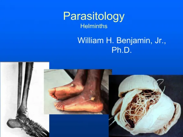
Elongated worm, cylindrical, unsegmented and tapering at both ends. Variable in size, measure <1 cm to about 100cm. Sex separate and male is smaller than female Nematodes:General features
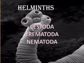
Nematodes: Location in the human body • Intestinal nematodes • Tissue nematodes
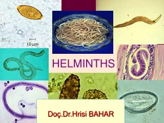
Enterobius(Oxyuris) vermicularis(Pinworm, seatworm, threadworm) Trichuristrichiura(whipworm) Ascarislumbricoides(roundworm) Ancylostomaduodenale & Necatoramericanus(hookworms) Strongyloidesstercoralis Nematodes: common intestinal infections
(Common names: Pin worm, seat worm( Found all over the world but more common in temperate regions. Children are more often evolved than adults, it tends to occur in groups living together such as families, army camps or nursery. Adult worms are mainly located in lumen of cecum and the female migrate to rectum to deposit her eggs on perianal skin. Direct human to human infection occurs mainly by swallowing the eggs. In addition, autoinfectionoccurs by contamination of the fingers. It can be seen by naked eye as white thread ± 1cm. Male is smaller than female ± 0.5cm, with coiled end. 1- Enterobiusvermicularis (THREAD WORM)
Pathology Majority of infections are asymptomatic. Main clinical presentation pruritus aniwhich can be very troublesome and occurs more often during the night, persistent itching may lead to inflammation and secondary bacterial infection of the peri-anal region. Infected children may suffer from emotional disturbance, insomnia, anorexia, loss of weight and loss of concentration and enuresis. Ectopic enterobiasis occurs in infected adult female when invade vulva and vagina result in valvo-vagintis, salpingitis, also adult worm can lodged in the lumen of appendix cause appendicitis. Enterobiusvermicularis (Oxyuris)
DIAGNOSIS : Unlike other intestinal Nematodes, the eggs are not usually found in feces.Thebest method is to look for them around the anus by taking an anal swab or by using CELLULOSE ADHESIVE TAPE the examination should be done before defecation or bathing. Treatment Albandazole or Mebendazolefor whole family Enterobiusvermicularis (Oxyuris)
The commonest human helminths infection all over the world. The large round worm which is normally located in the small intestine. Found in jejunumand upper part of ileum. Female ± 20 cm longer than male ± 10 cm Feed on semi digested food. Ascarislumbricoides (roundworm)
Ascarislumbricoides (roundworm) Infective stage is embryonated egg LIFE CYCLE
Ascaris eggs Ascaris larva emerging from egg Ascaris egg (embryonated)
Pathology: 1-Adult worm: (small intestine) Light infection : asymptomatic. Heavy infection : intestinal obstruction Migrating adult : to bile duct-jaundice 2-Larvae:Loeffler`s syndrome Pneumonitis and broncho-spasm, cough with bloody sputum Eosinophilia, urticaria Ascarislumbricoides (roundworm)
Ascarislumbricoides (roundworm) Loeffler`s syndrome:Larvae in lung pnuomonia, cough, bloody sputum
Ascarislumbricoides (roundworm) Ascaris larva in lung
Diagnosis: -eggs in stool. -larvae in sputum. -adult may pass with stool. Ascarislumbricoides (roundworm) Treatment: Albendazole or Mebendazole
Trichuris trichiura Infective stage is embryonated egg Diagnostic stage is egg in stool
World wide, common in poor sanitation. It coexists with Ascaris because of similar requirements (the eggs to be embryonated egg infective stage it needs to be 3 weeks in the soil). Adult live in large intestineespecially caecum and appendix–inheavy infection the whole length of large intestine affected. Male and female worm have narrowanterior portion penetrate the intestinal mucosa Trichuristrichiura(whipworm)
Pathology light infection: asymptomatic heavy infection: abdominal pain, bloody diarrhea. Rectal prolapsed in children is a common complication. Trichuristrichiura(Whipworm)
Diagnosis: egg in stool characterized by its barrel shape with mucoid plugs at each pole . Treatment: Albendazole. Trichuristrichiura(Whipworm)
Hook worms Ancylostomadudenale & Necatoramericanus Its buccal capsule (mouth) lined with hard hooks, triangular cutting plates and anticoagulant glands.
There are no specific symptoms or signs of hookworm infection, but they give rise to a combination of intestinal inflammation and progressive iron-deficiency anemia and protein deficiency Filariform Larval (infective stage) invasion of the skin can produce a skin disease called cutaneous larva migrans also known as creeping eruption, this is commonly caused by walking barefoot through areas contaminated with fecal matter. Larva migrate through the vascular system to the lungs, and from there up the trachea, and are swallowed. They then pass down the esophagus and enter the digestive system, finishing their journey in the smallintestinewhere the larvae mature into adult worms. They mate inside the host, females laying up to 30,000 eggs per day, which pass out in feces (diagnostic stage). The eggs need to be in soil for about one week to become FILARIFORM LARVA Buccal cavity attached to intestinal mucosa
Hook worms Pathology& clinical picture: • - larvae: • At the site of entry of larvae intense itching (ground itch) and dermatitis. • Migration phase: • cough with bloody sputum • pneumonitis and bronchitis but less sever than Ascaris, eosinophiliaurticaria. • - Adult worm: • low worm burden (INFECTION): no symptoms. • Moderate to heavy burden: • Epigastric pain, vomiting, hemorrhagic enteritis. • Protein loss: hypo-proteinaemia edema. • Anemia: due to withdrawal of blood by parasites and hemorrhage from punctured sites lead to sever anemia = microcytichypochromic anemia .
Diagnosis: -Eggs in stools.; -occult blood (+) Hook worms Diagnosis and treatment Treatment: Albendazol, Mebendazole
Strongyloidesstercoralis Widely distributed in tropical area at Asia, Africa & South America . Fatal dissemination in immuno-compromised host. It is smallest pathogenic nematodes ± 2.5mm. adult live in mucous membrane of duodenum jejunum rarely mucous membrane of bronchus. AUTOINFECTION IS VERY IMPORTANT CRITERIA .
Strongyloidesstercoralis life cycle • The parasite shows 3 different modes of development: • 1-Direct development: The rhabiditiform larva pass from stool and become directly a Filariform larva if the environment of the soil is suitable. • 2-Indirect development: in external environment Rh. larva becomes free living adults, produce eggs, rhabiditiform larva and Filariform larva(Infective stage). • 3-AUTOINFECTION: • Internal: when the rhabiditiform larva become a filariform larva in the intestine and penetrate the intestine • External: fecal contamination of skin –Rh larva > filariform penetrates the skin
Strongyloides stercoralis LIFE CYCLE
Cuteneouslittle reaction on penetration. sever dermatitis at perianal region in case of external autoinfection. Migration: pneumonitisduring larval migration. Intestinal: inflammation of upper intestinal mucosa, diarrhea, upper abdominal pain in the epigastria colicky in nature. Disseminated strongyloidiasis: in patient withimmunodeficiency, uncontrolled diarrhea –granulomatus changes –necrosis--perforation--peritonitis—death. Strongyloidesstercoralis: Pathology and clinical picture:
Diagnosis: rhabditiform larvae diagnostic stage in: -Stool examination -Duodenal aspirate Treatment:Albandazole, Mebendazole Strongyloidesstercoralis
DISEASE TRANSMISSION OF INFECTION LOCATION OF ADULT IN HUMANS LOCATION OF LARVA IN HUMANS CLINICALPICTURE Common Tapeworm Infections Taenia saginata taeniasis ingestion of larva in undercooked beef Small Intestine not present vague digestive disturbances eggs or proglottids in stools taeniasis ingestion of larva in undercooked pork Small Intestine not present vague digestive disturbances eggs or proglottids in stools Taenia solium- LARVA (cysticercus cellulosae) Cysticercosis ingestion of egg not present (except in Autoinfection, small intestine) sub-cutaneous muscles brain, eyes depending on locality: from none to epilepsy X-ray, CT, MRI Serology Hymenolepis nana hymenolepiais ingestion of egg Small Intestine Intestinal Villi Enteritis diarrhoea eggs in stools Echinochoccus granulosus hydatid disease ingestion of egg not present Liver, lungs, Bones etc depending on locality X-ray, CT, US Serology Hydatid sand LAB. DIAGNOSIS Taenia solium- ADULT TAPEWORM
Taeniasaginata • Is an obligatory parasite of man, the adult worm live in the SMALL INTESTINE. • CATTLE become infected by ingesting grass contaminated with eggs or gravid segments which passed from human faeces. In the cattle the onchosphere hatches out go to circulation and transformed to cysticercus stage in the muscle known as CYSTICERCUS BOVIS. • Man become infected by eating undercooked or improperly cooked beef, the adult worm lives in small intestine of man passing eggs and gravid proglottids to the environment. • The majority of cases are Asymptomatic, some patients have vague intestinal discomfort, vomiting and diarrhoea.
Location of hydatid cyst Echinococcus granulosus
Diagnosis of Hydatid cyst • Imaging: computed tomography (CT), magnetic resonance imaging (MRI) revealed a cystic swelling with smooth outline. • Microscopy: hyadtid sand • Serologic tests; to detect specific antibodies
Treatment of Tapeworms • Intestinal stages: Praziquantel • Tissue stages (Hydatid, cysticersosis): • Depends on clinical condition: Surgical and/or Albendazole