Download
1 / 28
280 likes | 503 Views
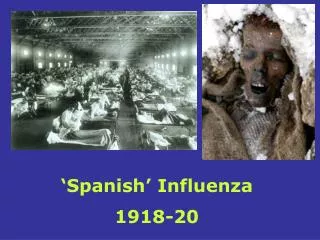
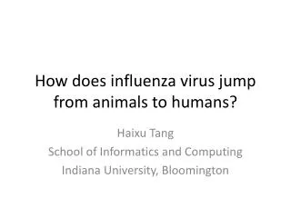
How does influenza virus jump from animals to humans?. Haixu Tang School of Informatics and Computing Indiana University, Bloomington. Swine flu outbreak, 2009. Influenza pandemics in human history. Spanish flu, 1918 Most deadly natural disaster Asian flu, 1957 Hong Kong flu, 1968

E N D
How does influenza virus jump from animals to humans? Haixu Tang School of Informatics and Computing Indiana University, Bloomington
Influenza pandemics in human history • Spanish flu, 1918 • Most deadly natural disaster • Asian flu, 1957 • Hong Kong flu, 1968 • Seasonal flu: every year • Vaccine designed based on the observation of the flu strains spreading in animals
Influenza is cause by a virus • Orthomyxoviridae; • a class of RNA virus using RNA as genetic material; • globular article of a diameter ~100 nm; • Protected by a bilayer and matrix proteins; • ~500 copies of H protein and ~100 copies of N proteins; • used for classification
Classification of influenza viruses • 16 H genes and 9 genes found • Spanish flu, 1918: H1N1 • Most deadly natural disaster • Asian flu, 1957: H2N2 • Hong Kong flu, 1968: H3N2 • Swine flu: 2009: H1N1 • Current pandemic threat: H5N1 • Several hundreds of active flu strains
Infection of flu viruses • Virus attached to the host cell; Host cell nucleus
Infection of flu viruses • Virus attached to the host cell; • Virus swallowed up by the host cell;
Infection of flu viruses • Virus is attached to the host cell; • Virus is swallowed up by the host cell; • Viral RNAs is released and enter the nucleus, where they are reproduced;
Infection of flu viruses • Virus is attached to the host cell; • Virus is swallowed up by the host cell; • Viral RNAs is released and enter the nucleus, where they are reproduced; • Fresh RNAs enter the cytosol;
Infection of flu viruses • Virus is attached to the host cell; • Virus is swallowed up by the host cell; • Viral RNAs is released and enter the nucleus, where they are reproduced; • Fresh RNAs enter the cytosol; • Viral RNAs act as mRNA to be translated into proteins forming new virus particles;
Infection of flu viruses • Virus is attached to the host cell; • Virus is swallowed up by the host cell; • Viral RNAs is released and enter the nucleus, where they are reproduced; • Fresh RNAs enter the cytosol; • Viral RNAs act as mRNA to be translated into proteins forming new virus particles; • New virus buds off from the membrane of the host cell;
Infection of flu viruses • Virus is attached to the host cell; • Virus is swallowed up by the host cell; • Viral RNAs is released and enter the nucleus, where they are reproduced; • Fresh RNAs enter the cytosol; • Viral RNAs act as mRNA to be translated into proteins forming new virus particles; • New virus buds off from the membrane of the host cell;
Spread of influenza viruses http://www.healtyhype.com
Recognition of the virus to the host cell:hemagglutinin (H protein) vs. glycans • On the surface of animal cells, there exist a heavy coat of glycans (sugars linked to proteins or lipids); • Different animals may have glycans of different structures; • Human, pig and birds • Different influenza virus strains with different hemagglutinin proteins (a class of glycan binding protein) recognize glycans of different structures
Structure of glycans linkage monosaccharide
Some basic graph theory • Graph: modeling pairwise relation (edges) between subjects (nodes or vertices) • Tree: a graph with no cycle • Each node in a tree has zero (leaves) or more child nodes • Subtree: a subset of nodes/edge • Labels • Nodes: monosaccharides • Edges: linkage types
Hemagglutinins recognize sialylated glycans • Human cells mainly express 2-6 linked sialylated glycans; • Bird cells mainly express 2-3 linked sialylated glycans; • Pig cells express both. Vessel theory
Hemagglutinin-glycan interaction is more complicated • Several influenza strains were observed to be inconsistent with the theory • AV18 strain has hemagglutinin proteins recognize specifically 2-3 linked glycans, but are not transmissable in birds; • NY18 and Tx91 strains recognize both 2-3 and 2-6 linked glycans, but NY18 does not transmit efficiently in human population, whereas Tx91 does; • Some chimeric H1N1 strains with high binding affinity to 2-6 linked glycans, but do not spread efficiently in human and pig.
Experimental determination of binding specificities of hemagglutinins Interaction assay hemagglutinin virus Glycan array The glycan motif finding problem
The glycan motif finding problem • Input: a set of (glycan) trees that are found to be recognized by a glycan binding protein (e.g. hemagglutinin) • Output: a l-treelet that is over-represented in the input set • l-treelet: a tree of a fixed small size l (e.g. l=4). • Over-representation: number of trees in the input set containing the treelet is much higher than the expected number in a random set of trees
Finding over-represented treelets Many 4-treelets appear in ALL input glycan trees. Glycans are not random!
The glycan motif finding problemA different formulation • Input: a positive set of (glycan) trees that are found to be recognized by a glycan binding protein (e.g. hemagglutinin) and a negative set of glycan trees that are found NOT to be recognized by the same protein • Output: a l-treelet that is over-represented in the input positive set than the negative set • Over-representation: number of trees in the positive set containing the treelet is much higher than the number of trees in the negative set
Fisher’s exact test: significance test of the over-represented treelets P=0.05 M glycans printed on the array N positive, M-N negative ni+ positives contain the treelet ni- negatives contain the treelet P=0.15
Glycan patterns found to be recognized by hemagglutinin • R. Sasisekharan and colleagues show is the pattern recognized by human influenza hemagglutinin, but not recognized by avian influenza hemagglutinin; • 2-6 linked sialylated glycans with long oligosaccharide branch (with multiple lactosamine repeats) are predominantly expressed in the human upper respratory epithelial cells • Indeed, the binding specificity of hemagglutinin to 2-6 linked sialylated glycans is not sufficient for the spread of the influenza viruses in human populations. Chandrasekaran A, et al. Nat Biotechnol , 2008; 26:107–113.
Spread of influenza viruses http://www.healtyhype.com