Download
1 / 31
360 likes | 676 Views
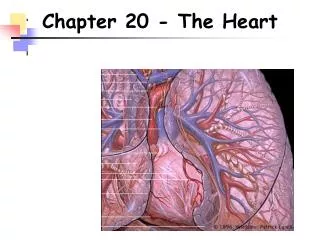
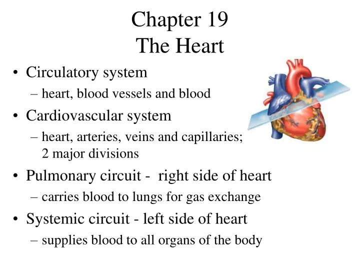
Chapter 19 The Heart. Circulatory system heart, blood vessels and blood Cardiovascular system heart, arteries, veins and capillaries; 2 major divisions Pulmonary circuit - right side of heart carries blood to lungs for gas exchange Systemic circuit - left side of heart

E N D
Chapter 19 The Heart • Circulatory system • heart, blood vessels and blood • Cardiovascular system • heart, arteries, veins and capillaries; 2 major divisions • Pulmonary circuit - right side of heart • carries blood to lungs for gas exchange • Systemic circuit - left side of heart • supplies blood to all organs of the body
Size, Shape and Position • Located in mediastinum, between lungs • Base - broad superior portion of heart • Apex - inferior end, tilts to the left, tapers to point • 3.5 in. wide at base, 5 in. from base to apex and 2.5 in. anterior to posterior; weighs 10 oz
Pericardium • Allows heart to beat without friction, room to expand and resists excessive expansion • Parietal pericardium • outer, tough, fibrous layer of CT • inner, thin, smooth, moist serous layer • Pericardial cavity • filled with pericardial fluid • Visceral pericardium (a.k.a. epicardium of heart wall) • thin, smooth, moist serous layer covers heart surface
Heart Wall • Epicardium (a.k.a. visceral pericardium) • serous membrane covers heart • Myocardium • thick muscular layer • fibrous skeleton - network of collagenous and elastic fibers • provides structural support • attachment for cardiac muscle • nonconductor important in coordinating contractile activity • Endocardium • smooth inner lining
Pericardium & Heart Wall • Pericardial cavity contains 5-30 ml of pericardial fluid
Heart Chambers • 4 chambers • Right and left atria • 2 superior, posterior chambers • receive blood returning to heart • Right and left ventricles • 2 inferior chambers • pump blood into arteries • Atrioventricular sulcus - separates atria, ventricles • Anterior and posterior sulci - grooves separate ventricles (next slide)
External Anatomy - Anterior Atrioventricular sulcus
Heart Chambers - Internal • Interatrial septum • wall that separates atria • Pectinate muscles • internal ridges of myocardium in right atrium and both auricles • Interventricular septum • wall that separates ventricles • Trabeculae carneae • internal ridges in both ventricles
Heart Internal Anatomy • Heart bisected in frontal plane, opened like a book
Heart Valves • Ensure one-way blood flow • Atrioventricular (AV) valves • right AV valve has 3 cusps (tricuspid valve) • left AV valve has 2 cusps (mitral,bicuspid valve) lamb • chordae tendineae - cords connect AV valves to papillary muscles (on floor of ventricles) • Semilunar valves - control flow into great arteries • pulmonary: from right ventricle into pulmonary trunk • aortic: from left ventricle into aorta
AV Valve Mechanics • Ventricles relax, pressure drops, semilunar valves close, AV valves open, blood flows from atria to ventricles • Ventricles contract, AV valves close(papillary m. contract and pull on chordae tendineae to prevent prolapse),pressure rises, semilunar valves open, blood flows into great vessels
Coronary Circulation • Blood vessels nourish cardiac muscle • Left coronary artery • anterior interventricular artery • supplies interventricular septum + anterior walls of ventricles • circumflex artery • passes around left side of heart in coronary sulcus, supplies left atrium and posterior wall of left ventricle • Right coronary artery • marginal artery • supplies lateral R atrium + ventricle • posterior interventricular artery • supplies posterior walls of ventricles
Venous Drainage • 20% drains directly into right ventricle • 80% returns to right atrium • great cardiac vein (anterior interventricular sulcus) • middle cardiac vein (posterior sulcus) • coronary sinus (posterior coronary sulcus) collects blood from these and smaller veins and empties into right atrium
Coronary Flow and the Cardiac Cycle • Reduced during ventricular contraction • arteries compressed • Increased during ventricular relaxation • openings to coronary arteries, just above aortic semilunar valve, fill as blood surges back to valve
Metabolism of Cardiac Muscle • Aerobic respiration • Rich in myoglobin and glycogen • Large mitochondria • Organic fuels: fatty acids, glucose, ketones • Fatigue resistant