Download
1 / 23
230 likes | 434 Views
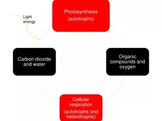
Energy Releasing Pathways (Cell Respiration). I . Introduction. A. History. 1. Antoine Lavoisier in the 1700’s can make wine without living organisms. 2. Wohler and von Leibig supported this idea, but Schwann showed juice would not ferment without yeast.

E N D
Energy Releasing Pathways (Cell Respiration) I. Introduction A. History 1. Antoine Lavoisier in the 1700’s can make wine without living organisms 2. Wohler and von Leibig supported this idea, but Schwann showed juice would not ferment without yeast. 3. In 1860 Pasteur proved ethanol amount proportional to the amount of yeast present
4. In 1897 the Buchner brothers steps of glycolysis key to fermentation 5. In the early 1900’s Szent-Gyorgyi designed Citric Acid Cycle, failed to show relationship to fermentation 6. Krebs in 1938 linked glycolysis to citric Acid Cycle via enzyme CoA Kreb’s Cycle
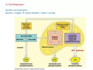
II. Aerobic Respiration A. Overview Figure 9.6
B. Glycolysis 1. Where occurs? a.Cytosol Figure 6.9 Figure 6.9
2. Steps and Players a. Components: i. Investment, & iii. Harvest ii. Splitting, Figure 9.9
i. Investment 1. Kinase enzyme attaches a P from ATP to glucose (6C) making glucose-P Prevents glucose from moving back out of cell 2. Isomerase rearranges glucose-P into fructose-P (6C) Prepares molecule to add another Phosphate 3. Kinase enzyme attaches another P from second ATP to fructose-P, making P-fructose-P Generates a balanced molecule with a P at either end. ii. Splitting 1. Aldolase enzyme cuts molecule P-fructose-P into two 3C molecules G3P and Dihydroxyacetone-P 2. Dehydrogenase enzyme liberates H and NAD+ steals the electrons from H This happens twice or once for each G3P 3. The hole left by the leaving H is backfilled by Pi and forms G1,3P This step balances the two G3P’s with a P on both ends How many NADH + H are formed per glucose?
iii. Harvest 1. Kinase enzyme directly transfers a P from G3P to ADP to make ATP by substrate level phosphorylation (SLP) How many times does this happen to make how many ATP’s? 2. Mutase enzyme rearranges G3P into G2P Prepares molecule for more harvest 3. Enolase enzyme rearranges G2P into PEP Prepares molecule for more harvest 4. Kinase enzyme directly transfers a P from PEP to ADP to make ATP by SLP Makes pyruvate out of each PEP
3. Outcomes a. 2ATP are used by the cell. The next two outcomes only happen if oxygen is present in the cell. b. NADH + H mitochondria and electron transport chain c. 2pyruvic acids are combined to CoA to go to the mitochondria and the Kreb’s cycle
C. Transport 1. Where occurs? a. Cytoplasm to Mitochondria Figure 9.10 2. Steps How many times this happen? a. Dehydrogenase enzymes splits off a CO2 from pyruvic acid which liberates electrons from H and given to NAD+ to make a 2C acetyl group b. Combine acetyl group to Co-enzyme A to be transported to the mitochondria
3. Outcomes The next three outcomes only happen if oxygen is present in the cell. a. NADH + H mitochondria and electron transport chain b. 2pyruvic acids combined to 2CoA go to the mitochondria and the Kreb’s cycle c. What is the fate of the CO2?
D. Krebs Cycle 1. Where occurs? a. Mitochondrial Matrix Figure 6.17
2. Steps a. Divisions i. Destroying ii. Rearranging Figure 9.12
i. Destroying 1. Enzyme combines acetic group to oxaloacetic acid to begin cycle 2. Dehydrogenase enzymes splits out CO2 and liberates H to NAD+ How many CO2 are liberated? 3. As H’s are removed then a Pi jumps on only to be removed to form ATP by SLP ii. Rearranging 1. Mutase and dehydrogenase enzymes reshape molecule to liberate more H’s to rebuild oxaloacetic acid 2. Liberates H and NAD+ or FAD steals the electrons This happens twice for glucose or once for each acetic group.
3. Outcomes c. NADH + H and FADH2 are sent to electron transport chain a. ATP is used b. CO2 diffuses into cytosol and is lost
E. Electron Transport Chain 1. Where occurs? a. Inner Mitochondrial Membrane Figure 6.17
2. Steps a. Divisions i. Build-up & ii. Harvest Figure 9.13 Figure 9.14
i. Build Up 1. NADH and FADH2 drop the electrons from H to a series of re-dox proteins called cytochromes 2. As electrons move down the chain they lose energy which is used to move the H proton across the membrane to establish potential energy ii. Harvest 1. The electrons are eventually passed to an awaiting Oxygen atom 2. The H proton moves back across the membrane through ATP Synthase and to the waiting O2 to form water 3. Conversion of energy (Potential to Kinetic) is used to form ATP
3. Outcomes b. NAD+ and FAD+ sent back to glycolysis or the Kreb’s cycle a. ATP is used c. Water moved out or used
Summary of Aerobic Respiration Figure 9.16
III. Anaerobic Respiration A. Fermentation 2. Process 1. Who? only glycolysis Figure 9.17a
A. Lactic Acid Shuttle 1. Who? 2. Process Animal cells == lactic acid shuttle and Liver Figure 9.17b
IV. Versatility A. Routes B. Problems Figure 9.19
V. Regulation A. Mechanisms B. Sites Figure 9.20