Download
1 / 38
1.04k likes | 3.88k Views
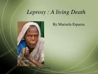
Leprosy Hansen’s disease. Introduction . Leprosy is a chronic granulomatous infection of the skin and the peripheral nerves, with an intracellular bacterium Mycobacterium Lepra.

E N D
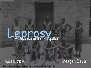
Leprosy Hansen’s disease
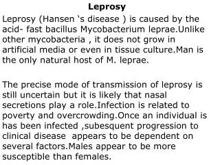
Introduction • Leprosy is a chronic granulomatous infection of the skin and the peripheral nerves, with an intracellular bacterium Mycobacterium Lepra. • Leprosy was recognized in the ancient civilization of China, Egypt, and India. The first known written mention of leprosy is dated 600BC.
Introduction • Leprosy shows a wide range of clinical presentation from: • Tuberculoid leprosy (TT) • Borderline leprosy: borderline tuberculoid (BT), midborderline (BB), borderline lepromatous (BL). • Lepromatous leprosy (LL). (Classified by Ridley & Jopling 1966)
A simpler field classifications depending on the number of skin lesions: • Single skin lesion (one patch). • Paucibacillary ( 2-5 patchs). • Multibacillary more than 5 patchs)
Etiologic agent • Mycobacterium Leprae, discovered in 1873 by G.A. Hansen. • Intracellular parasite with tropism for macrophages and Schwann cells. • Viable bacilli stained with carbol-fuchsin appear as solid rods with rounded ends, while those is irregular stain are dead.
Etiologic agent • M. leprae is an acid & alcohol fast (Ziehl-Neelsen), gram positive bacilli. • Prefer to grow in cooler regions of the body < 37ºc. • It has never been cultivated extracellularly, but organism can replicate in mouse footpad & 9-banded armadillo
Etiologic agent • Genome include 1605 genes encoding proteins & 50 genes for stable RNA molecule. • More than half of the functional genes are replaced by inactive or pseudogenes, retaining the genes essential for Mycobacterial cell wall formation. • Thus M. leprae depend on host metabolic products, and this explain it’s slow rate of replication and inability to grow in culture. (Shin Y,et al. 2000)
Etiologic agent • Mycobacterial cell wall contain several targets for host immune response: • Phenolic glycolipid I (PGL-I). • Lipoarabinomannan. • Other cell wall proteins purified from glycolipid component of cell wall.
Phenolic glycolipid I (PGL-I). • Prominent surface lipid specific for M. leprae. • Best characterized virulent factor: • Binds to C3,»»» phagocytosis of the bacterium by mononuclear phagocytes. • Protect against oxidative killing by hydroxyl radicals and superoxide anions.
Phenolic glycolipid I (PGL-I). • Specific tropism for Schwann cells: • Trisaccharide terminal of PGL-I »»» G domain of α 2 chain of laminin 2, a basal component of lamina of Schwann cells restricted to peripheral nerves. • Stimulates host immune response: • Potent IgM antibody response, that is proportional to bacterial load and fall with therapy. (Sridharan, et al. 2005)
Lipoarabinomannan: • Modulates macrophages phagocytic activity, & proteins involved in cell wall synthesis. • Other cell wall proteins purified from glycolipid component of cell wall: • Act as potent T-cell antigens, stimulate protective immunity in murine M. leprae infection. (Britton & Lockwood, 2004)
Epidemiology Prevalence: • In the past 20 years more than 14 million patients have been cured. • In 1985 »»» 12/10 000, dropped in 2000 »» > 1/10 000, with a 20% annual decrease in new cases detected globally since 2001. • Leprosy is eliminated from 113 countries of 122 where leprosy was considered a public health problem in 1985.
Epidemiology • In 9 countries in Africa, Asia & Latin America »»» more than 1/10 000 • Eighty three % of cases are present in 6 countries: India, Brazil, Burma, Indonesia, Madagascar & Nepal. • India account for 64% of cases world wide. (WHO, 2005)
WHO African region leprosy elimination program meeting in 2003
Epidemiology • Primary host is human. Naturally occurring infection is also reported armadillos in America & African chimpanzee. • Sex: Male more affected than females (M/F ratio 1.5-2 to 1) except in some areas of Africa. • Age: All ages, about 20% of cases occur in children below 10 years, but it is extremely rare in infants.
Transmission • Aerosol spread of nasal secretion, & uptake through nasal or respiratory mucosa. • M. Leprae in nasal secretion can survive up to 36 hours, or as much as nine days in tropical areas. • Proximity is an important determinant of transmission, & incidence among houses hold contacts: 8-10 for lepromatous leprosy. 2-4 for tuberculoid leprosy (Sridharan, et al. 2005)
Incubation Period • Varies widely from months to 30 years • With a mean of 4 years for TT and 10 years for LL. • Subclinical infection is reported in areas of high prevalence, as M. leprae DNA was detected in 5% of nasal swabs from normal individuals. (Britton & Lockwood, 2004)
Host susceptibility • Genetic factors can affect both the development and the pattern of the disease: • susceptibility loci on chromosomes 10 & 6 »» Indian patients. • Polymorphism TNF- promoter genes »» multibacillary leprosy in Brazilian patients. • HLA DR2 & DR3 »» »» tuberculoid diseases, while HLA DQ1with lepromatous form • Mutation in toll-like receptor-2 gene »» »» lepromatous leprosy in Koreans. (Britton & Lockwood, 2004)
Pathogenesis • Clinical forms of the diseases depend on the ability to mount a cell mediated immune response. • One pole (TT) »»vigorous CMI »» lesions infiltrated with Th1-like Tcell» » IFN , TNF α, IL-2 and IL-15 »»well demarcated granulomas with few mycobacteria found in the lesions. • Diseases is limited to few well defined skin patchs or nerve trunks.
Other pole (LL): • Absent specific cellular immunity,»» uncontrolled proliferation of bacilli with many lesions and extensive infiltration of the skin and nerves. • There is no organized granuloma, foamy macrophages, and high antibody titer to PGL-I. • Deviation of CD4+ve T cells, with Th2 like cytokines IL-4 and IL-10, deletion of T cells, or suppressor T cells
Pathogenesis • Most of the patients have the intermediate form (BT,BB, and BL). • They are unstable and either progress: • Slowly toward lepromatous pole, or • Type 1 leprosy reaction (reversal reaction) Spontaneous increased T cell reactivity & in cytokines IFN , TNF α • Type 2 reaction (erythema nodosum leprosum) Systemic inflammatory immune response»» extravascular immune complexes »» neutrophil infiltration, complement activation and high concentration of TNF α
Clinical features • Skin involvement: Commonly macules or plaques rarely papules or nodules are seen. In tuberculoid and BT, lesions are few, hypopigmented with raised edges, and with reduced sensation
Clinical features • Lepromatous form, many skin lesion, symmetrical, confluent in some cases, and many of them are not hypoaesthetic.
Clinical features • Nerve damage: • Peripheral nerve trunk damage: Posterior tibial, ulnar, median, lateral popliteal and facial. Involved nerves are enlarged, and with regional sensory and motor loss. • Small dermal sensory & autonomic nerves: Hypoaesthesia »» TT and BT. Glove & stocking »» lepromatous form. • Pure neuritic leprosy:
Clinical features • Eye involvement: • Blindness »»» nerve damage & direct invasion • Lagophthalmus »»» orbicularis oculi »»» zygomatic & temporal branches of facial nerve. • Corneal ulceration »»» anaesthesia »»» ophthalmic branch of trigeminal nerve.
Clinical features • Systemic features: • Nasal mucosa »»» cartilage »»» saddle shape. • Bone destruction »»» osteomylitis. • Testicular atrophy »»» loss of testosterone . • Renal involvement and amyloidosis
Diagnosis • Lepromin test: • Intradermal inoculation of killed M leprae. • Early reactions (48 h, Fernandez) • Late reactions (3-4 wk, Mitsuda) • strongly positive responses (>5 mm) in TT or BT, while patients with LL do not respond.
Diagnosis • Slit smear technique: • Skin incision in 6 sites ( ear lobes, elbow, knee and a lesion) • Slit is smeared on a slide and stained with Ziehl-Neelsen. • Microscopic score (1+ to 6+), reflect number of bacilli by HPF • Useful to quantitate bacterial load • High specificity but low sensitivity 70%
Diagnosis • Serology: • ELISA: to detect antibodies against carbohydrate portion of the PGL-I. • Postive in lepromatous but not the tuberculoid form. • Antibody titer decreases with effective therapy. • Polymarase chain reaction (PCR): • Amplify the DNA of M leprae • Low bacterial loads (<10 bacilli) can be detected. • 60-75% of smear -ve patients with TT leprosy have positive results on PCR.
Diagnosis • Histologic diagnosis: • In TT: Noncaseating granuloma, bacilli are few or absent, dermal nerve involvement, with normal skin organs. • In LL: Diffuse granulomatous reaction, foamy macrophages, more common around blood vessels and nerves
Treatment • Chemotherapy: All patient should receive multi-drug therapy (MDT). First line drugs: Rifampicin, Clofazimine, and Dapsone.
Treatment Second line therapy: Minocycline, clarithromycin, and ofloxacin, are highly effective against M. leprae. Reversal reaction: Peak time: during the first 2 month of therapy, even up 12 months, and after (MDT) is completed. Corticosteroids 40-60mg daily, taper 5 mg every 2-4 weeks, duration of therapy 3-4 months. Recovery rate for nerve function 60-70%, less with pre-existing nerve damage or recurrent reaction.
Treatment Type 2 reaction (ENL): Develop during 1st or 2nd year of MDT, and can relapse over several years. Anti- inflammatory: Clofazimine 300mg daily. Or Drug that target overproduction of TNF-α,Thalidomide 400mg daily, or pentoxifylline. Or Neutralization of TNF-α with monoclonal antibodies, or soluble inhibitors.
Prophylaxis • Immunoprophylaxis: • BCG offer variable protection against leprosy (34-80%) in different countries, adding heat –killed M. leprae increases the protective effect to 64%. • Endemicity of leprosy, background saprophytic mycobacterial flora, and the age at vaccine may affect the response to vaccination. • Vaccination may precipitate TT leprosy in apparently healthy contacts, thus immunoprophylaxis is best carried out at an early age. • Chemoprophylaxis: • Rifampicin, to close contact of a case, and can be give to children under the age 12 years (15mg/kg monthly for 6 months)
Pregnancy & leprosy • Little evidence that pregnancy can cause new diseases or relapse. • However, pregnant BL patients may experience type 1 reaction in the post partum period. • Also lepromatous leprosy patients can experience ENL during pregnancy and lactation, with early loss of nerve function than non-pregnant patients. • MDT (Rifampicin, Clofazimine, and Dapsone) is safe.
HIV infection & leprosy • Unlike tuberculosis, leprosy is not significantly associated with HIV infection. • HIV-associated neuropathy might be confused with leprosy neuritis, also neuropathy due to antiretroviral chemotherapy might be confused with leprosy. • There is difference in treatment strategy for patients with leprosy and HIV, icluding treatment of reactions.


![[PDF] Free Download First Man By James R. Hansen](https://cdn4.slideserve.com/8039496/slide1-dt.jpg)