Download
1 / 1
10 likes | 165 Views
Rodrigo França de Espíndola , MD 1 ; Marcony Rodrigues Santhiago , MD 1,2 ; Paula de Camargo Abou Mourad , MD 1 ; Marco Aurélio Costa Marcondes, MD 1 ; Flávio Gaieta Holzchuh, MD 1 ; Mário Luiz Ribeiro Monteiro MD, PhD 1 ; Newton Kara-Junior , MD,PhD 1

E N D
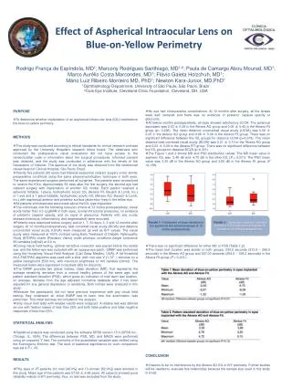
Rodrigo França de Espíndola, MD1; Marcony Rodrigues Santhiago, MD1,2; Paula de Camargo Abou Mourad, MD1; Marco Aurélio Costa Marcondes, MD1; Flávio Gaieta Holzchuh, MD1; Mário Luiz Ribeiro Monteiro MD, PhD1; Newton Kara-Junior, MD,PhD1 1Ophthalmology Department, University of São Paulo, São Paulo, Brazil 2Cole Eye Institute, Cleveland Clinic Foundation, Cleveland, OH, USA • PURPOSE • To determine whether implantation of an aspherical intraocular lens (IOL) interference the blue-on-yellow perimetry. • METHODS • This study was conducted according to ethical standards for clinical research and was approved by the University Hospital’s research ethics board. The observers who conducted the postoperative visual evaluations did not have access to the randomization code or information about the surgical procedures. Informed consent was obtained, and the study was conducted in adherence with the tenets of the Declaration of Helsinki. The approval of the study was obtained from the institutional review board of Clinical Hospital, São Paulo, Brazil • Twenty-five patients (50 eyes) had bilateral sequential cataract surgery under similar preoperative conditions using the same phacoemulsification technique in both eyes. The same experienced surgeon performed all surgeries. The patients were randomized to receive the IOLs. Approximately 30 days after the first surgery, the second eye had cataract surgery with implantation of another IOL model. Each patient received a spherical foldable, 1-piece, hydrophobic acrylic IOL (Akreos Fit, Bausch & Lomb, Inc.) in 1 eye and a 1-piece foldable, hydrophobic acrylic IOL (Akreos AO, Bausch & Lomb, Inc.) with aspherical anterior and posterior surface (aberration free) in the fellow eye. • All patients and observers were mask about the IOL type implanted. • The individuals met the following inclusion criteria at 12 moths postoperatively: visual acuity better than 0.3 LogMAR in both eyes, normal intraocular pressures, no evidence of posterior capsule opacity, and no signs of glaucoma. Patients with any ocular disease (infectious, inflammatory, and degenerative) were excluded. • Patients were examined before surgery and at 1, 7, 30 days, 3, 6 and 12 months after surgery. At 12 months postoperatively, best-corrected visual acuity (BCVA) and distance uncorrected visual acuity (UCVA) were measured as well as B/Y values. The visual acuity were measured at 100% contrast using Early Treatment of Diabetic Retinopathy Study (ETDRS) charts (Precision Vision) under photopic conditions (target luminance 85 candelas [cd]/m2) at 4.0 m. • During visual field testing, optimal refractive correction was placed before the tested eye, and the fellow eye was occluded with an opaque eye patch. SWAP was performed using the Humphrey Visual Field Analyzer (Carl ZeissMeditec, USA). A full-threshold 24-2 FASTPAC algorithm was used with a blue (440 nm) size V (1.72°) stimulus on a yellow background (530 nm), with maximum brightness of 100 candela (cd/m2). The measured levels were expressed in decibels (dB) for all points. • The SWAP provides two global indices; mean deviation (MD), that represents the average sensitivity deviation from a normal healthy person of the same age; and pattern standard deviation (PSD), which gives an indication of how each test location, on average, deviates from the age adjusted normative database after it has been adjusted for any general depression or sensitivity. Both indices were analyzed in this study • Because the participants did not have previous experience with any visual field testing, they underwent an initial SWAP test to learn how the examination was performed. This initial test was not included in the analysis. • Only visual field tests with reliable results were analyzed. A reliable test was defined as one with fixation losses of less than 25% and both false-positive and false-negative responses of less than 33%. • STATISTICAL ANALYSIS • Statistical analysis was conducted using the software SPSS version 15.0 (SPSS Inc., Chicago, IL, USA). The differences between PSD, MD, and MACS were performed using an unpaired “t” test. The normality of the quantitative variables was verified using the Kolmogorov-Smirnov test. The level of statistical significance for such comparison was set a P <. 05. • RESULTS • Fifty eyes of 25 patients (12 men [48.0%] and 13 women [52.0%]) were enrolled in this study. Mean age of the patients was 57.80 ± 6.48 years. Allsubjectsshowedgoodreliability indices in B/Y perimetry, thus, no test was excluded from the study. • No eye had intraoperative complications. At 12 months after surgery, all the lenses were well centered and there was no evidence of posterior capsule opacity or glaucoma. • At twelve months postoperatively, all eyes showed satisfactory UCVA. The spherical equivalent was 0.03 ± 0.29 in the Akreos AO group and 0.06 ± 0.43 in the Akreos Fit group (p= 0.296). The mean distance uncorrected visual acuity (UCVA) was 0.08 ± 0.05 in the Akreos AO group and 0.09 ± 0.06 in the Akreos Fit group. There was no significant difference between the IOL groups for distance UCVA (p=0.379). The mean distance best corrected visual acuity (BCVA) was 0.01 ± 0.10 in the Akreos AO group and 0.02 ± 0.09 in the Akreos FIT group. There was no significant difference between the IOL groups for distance BCVA (p=0.331). • TheFigure 1 and 2 shows MD and PSD distribuitionvalues. The MD meanvalue in aspheric IOL was -2,40 dB and -4,70 dB in theother IOL (P = 0,071). The PSD meanvaluewas 3,30 dB in theAkreos AO groupand 3,80 dB in theAkreosFitgroup (P =0,136). • Therewas no significantdifference for either MD or PSD (Table 1,2). • The mean test duration was similar in both groups, 230,5 seconds (213,5 – 266,0 seconds) in theAkreos AO groupand 227,00 seconds (204,0 – 324,2 seconds) in theAkreosFitgroup (P = 0,213.). • CONCLUSION • It seems to be no interference by the Akreos AO IOL in B/Y perimetry. Further studies will be needed to evaluate this relationship because the sample size used in this study is small. Effectof AsphericalIntraocularLensonBlue-on-YellowPerimetry Figure 1 -Distributionof meandeviation in theasphericalandsphericalgroupsat 12 mothspostoperatively. Figure 1 -Distribution of pattern standard deviation in theasphericalandsphericalgroupsat 12 mothspostoperatively.