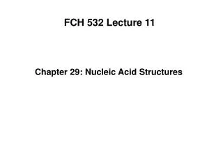
FCH 532 Lecture 14
310 likes | 597 Views
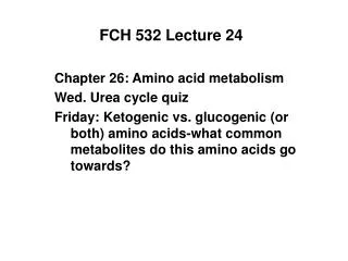
FCH 532 Lecture 14. Quiz Friday on nucleic acid structures, base pairing Extra credit assignment for Friday March 2-Seminar Speaker Matt DeLisa Chapter 30. SOS response. SOS response causes cells to stop dividing and repair damaged DNA. LexA and RecA mutants always have the SOS response on.

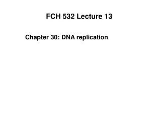
FCH 532 Lecture 14
E N D
Presentation Transcript
FCH 532 Lecture 14 Quiz Friday on nucleic acid structures, base pairing Extra credit assignment for Friday March 2-Seminar Speaker Matt DeLisa Chapter 30
SOS response • SOS response causes cells to stop dividing and repair damaged DNA. • LexA and RecA mutants always have the SOS response on. • When E. coli is exposed to agents that damage DNA, RecA mediates proteolytic cleavage of LexA. This is induced by RecA binding to ssDNA. • LexA is a repressor of 43 genes involved in DNA repair (all proceeded by 20 nt sequence called the SOS box).
Figure 30-59 Regulation of the SOS response in E. coli. Page 1180
SOS Repair • E. coli Pol III is unable to replicate through lesions (AP sites, thymine dimers), causing a replication fork “collapse” • To restore the replication fork, can either induce recombination repair which uses a homologous chromosome as the template or SOS repair. • Uses 2 bypass DNA polymreases (Pol IV and PolV). • These are error-prone DNA polymerases (lack the 3’ 5’ exonuclease) • SOS is a mutagenic process. This is a last resort if DNA has not been repaired by other mechanisms.
Double-strand break (DSB) repair • Double-strand breaks (DSBs) in DNA are produced by ionizing radiation and free radical products of oxidative metabolism. • Can also occur as intermediates in meiosis. • Unrepaired DSBs can lead to cancer or cell death. • 2 modes to repair DSBs • 1. Recombination repair • 2. Nonhomologous end-joining (NHEJ)
Nonhomologous end-joining (NHEJ) • Broken ends are aligned and frayed ends are trimmed or filled in, and their strands ligated. • NHEJ in eukaryotes has several proteins: • Ku-heterodimer of Ku70 and Ku80 • DNA ligase IV • Xrcc4 • Ku binds to double-stranded breaks binding to DNA’s major and minor grooves. • Nucleotide trimming is carried out by ATP-dependent Mre11 complex. • Gaps filled in by DNA polymerases and sealed by DNA ligase IV and Xrcc4.
Figure 30-62 Schematic diagram of nonhomologous end-joining (NHEJ). Page 1183
Carcinogens • Carcinogens-chemical agents that can cause cancer • Up to 80% caused by exposure to these agents • Usually damage DNA; likely to induce SOS response, so they are indirect mutagenic agents. • High correlation between mutagenesis and carcinogenesis. • Ames Test-assays for carcinogenicity • Salmonella typhimurium his- strain- must be grown in presence of His. • Lack lipopolysaccaride coats and are highly permeable to many substances. • Inactivated excision repair systems. • Look for reversion to his+ phenotype.
Ames Test • ~109 cells spread on plate lacking His. • Use a mixture of his- strains to detect base changes, insertions and deletions. • Mutagen is place in culture medium that causes some his- strains to revert to his+ • Mutagenicity is scored as # of colonies less the few spontaneously revertant colonies that occur in the absence of the mutagen. • Many noncarcinogens are converted to carcinogens in liver. • Small amount of rat liver is added to Ames test to mimic mammalian metaboilsm.
Homologous recombination • Defined as the exchange of homologous segments between two DNA molecules. • Bacteria are haploid and acquire foreign DNA through conjugation in which DNA is directly transferred from one cell to another via a cytoplasmic bridge. • Holliday junction - corresponding strands of two aligned homologous DNA duplexes are nicked and the nicked strands cross over to pair with the nearly complementary strands of the homologous duplex after which the nicks are sealed.
Figure 30-64 The Holliday model of homologous recombination between homologous DNA duplexes. Page 1184
Formation of Holliday Junction Branch migration-4 strands exchange base pairing partners Page 1184
Resolution of Holliday Junction occurs in 2 ways The cleavage of the strands that did not cross over exchanges the ends of the original duplexes to form, after nick sealing, traditional recombinant DNA 2. The cleavage of strands that crossed over exchanges a pair of homologous single-stranded segments.
Figure 30-65a Secondary structure of the four-stranded Holliday junction of self-complementary decameric DNA d(CCGGTACCGG). Watson-Crick base pairing interactions are shown in black. 2-fold axis relating two helices of stacked-X conformation is represented by large black lenticular symbol. Page 1185
X-ray structure of the cross over event. Page 1185
Figure 30-66 Homologous recombination between two circular DNA duplexes. This process can result either in two circles of the original sizes or in a single composite circle. Page 1186
Figure 30-67a Electron micrographs of intermediates in the homologous recombination of two plasmids. (a) A figure-8 structure. This corresponds to Fig. 30-66d. Page 1186
Figure 30-67b Electron micrographs of intermediates in the homologous recombination of two plasmids. (b) A chi structure that results from the treatment of a figure-8 structure with a restriction endonuclease. Page 1186
Homologous recombination catalyzed by RecA in E. coli • recA-E.coli have 104-fold lower recombination rate than wild-type cells. • RecA polymerizes cooperatively on ssDNA or dsDNA that has a single-stranded gap. • Resulting filaments may contain 1000s of RecA molecules. • Bind homologous dsDNA and using ATP catalyze strand exchange.
Figure 30-68 An electron microscopy–based image (transparent surface) of an E. coli RecA–dsDNA–ATP filament. Page 1187
Figure 30-69a X-Ray structure of E. coli RecA protein. Monomers yellow and blue, ADPs red. (a) View normal to the helix axis as in Fig. 30-68. Page 1187
Figure 30-69b X-Ray structure of E. coli RecA protein. (b) View nearly parallel to the helix axis showing one turn of the helix. Page 1187
Figure 30-70 A model for RecA-mediated pairing and strand exchange between a single-stranded and a duplex DNA. ssDNA binds to RecA to form initiation complex. dsDNA binds to the initiation complex to form a transient 3-stranded helix to mediate correct pairing to homologous strand. RecA rotates the bases of the aligned homologousstrands to effect strand exchange in an ATP-driven process. Page 1188
Figure 30-71 The RecA-catalyzed assimilation of a single-stranded circle by a dsDNA can occur only if the dsDNA has a 3¢ end that can base pair with the circle (red strand). Page 1188
Figure 30-72 A hypothetical model for the RecA-mediated strand exchange reaction. Page 1189
Recombination repair • Damaged replication forks occur at a frequency of at least once per bacterial cell generation and 10 X per eukaryotic cell cycle. • DNA lesions that damage replication forks can be repaired through recombination repair. • Happens when a replication fork encounters an unrepaired single-strand lesion.
Figure 30-77 The recombination repair for a single-strand lesion. DNA replication is stopped at the lesion but continues on the opposing undamaged strand before replisome collapses. Replication fork changes to a Holliday junction (Chicken Foot). Single-strand gap at collapsed replication fork now an overhang is filled in by Pol I Reverse branch migration mediated by RuvAB or RecG yields a reconstituted replication fork. Page 1192
Figure 30-78 The recombination repair for a single-strand nick. Single stranded nick causes replication fork to collapse. Repair process: RecBCD and RecA invasion of newly synthesized and undamaged 3’-ending strand into homologous dsDNA Branch migration via RuvAB makes Holliday junction to exchange 3’-ending strands. RuvC resolves Hollidayh junction making the 5’ end strand nick becomes a 5’ endo fo Okazaki fragment. Page 1193
Figure 30-79 The repair DSB in DNA by homologous end-joining. DSB double-stranded ends are changed to single-stranded ends. The 3’-ending strands pairs invades the homologous chromosme to form a pair of Holliday junctions. DNA synthesis and ligation to fill gaps and seal Both Holliday junctions are resolved. Page 1193