Download
1 / 14
160 likes | 366 Views
The Role of the Basal Ganglia in Habit Formation By Henry H. Yin & Barbara J. Knowlton. Group 3, Week 10. Alicia Iafonaro Kimberly Villalva Tawni Voyles. 1. Discuss the relationship of BG and hippocampus to place and response strategies.

E N D
The Role of the Basal Ganglia in Habit Formation By Henry H. Yin & Barbara J. Knowlton Group 3, Week 10 Alicia Iafonaro Kimberly Villalva Tawni Voyles
1. Discuss the relationship of BG and hippocampus to place and response strategies. Poldrack and Packard argued that direct or indirect neural connections between the hippocampus and dorsal striatum could mediate the competition between them. Tolman, Packard, and McGaugh- trained rats to retrieve food from one arm of a cross maze surrounded by various environmental cues. • After training it is given a probe test- starting arm is placed at the opposite end of the maze. • If the rat used the response strategy and turned left, shows that the learning was inflexible and response-specific. If the rat used the place strategy and turned right, shows that the animal was able to in corporate surrounding spatial cues in deciding (choose opposite of what was learned in training).
1. Discuss the relationship of BG and hippocampus to place and response strategies. Results • Most rats used the place strategy during the probe test, but after extensive training they switched to a response strategy. • Rats were more likely to use the place strategy despite extended training showed inactivity of the dorsal striatum. • Rats who used the response strategy more frequently, even early in training showed inactivity of the hippocampus. Support for Poldrack and Packard idea of the dorsal striatum and the hippocampus can be viewed as competing learning systems because each strategy used inactivity in either the dorsal striatum or hippocampus.
1. Discuss the relationship of BG and hippocampus to place and response strategies. The authors suggest that the hippocampus does not compete with or function independently of the dorsal striatum. They can act together with other regions, such as the medial and ventral striatal regions form a function circuit. • DSM contains special selective neurons that fire when animals take a particular route to reach a goal. • Contains head- direction neurons with activity aligned with that of the place fields of hippocampal place cells. • Information about current position provided by these cells can be used to signal where to go to reach a definite goal. • This information is suggest that is information is conveyed to the DMS via the cortico-striatal projection from the hippocampal pyramidal neurons.
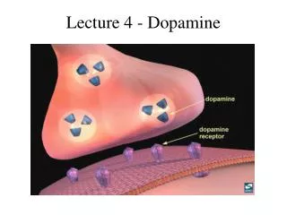
In the Probabilistic Clarification Task: 2. How does performance learning in different tasks tell us something about the different functions of the two learning systems? . . . • Patients w/ Amnesia: • Learned associations normally, independently of MTL structures that support declarative memory. • Assumed to rely on non-declarative learning. • Patients w/ Parkinson's: • These patients had abnormal striatal functioning due to loss of dopaminergic input. • Impaired in the implicit learning of these associations, although they achieved normal levels of performance with further training • MRI: showed activation in the hippocampus & surrounding MTL cortical regions. • Achieved good performance by relying on declarative memory. There is a dissociation between declarative learning (dependent on the MTL) and non-declarative (habit) learning (dependent on striatum). • Control Participants: • Activation in striatal regions during learning. • Relied on non-declarative learning mechanisms.
. . . Are the two systems always operating in a mutually exclusive way? Results suggest that multiple neural systems can support learning in the probabilistic clarification task. Many real world tasks encountered by humans probably involve BOTH habit and declarative learning. The system that contributes most to performance depends on: The amount of training The ease of memorizing associations between stimuli The relative integrity of the basal ganglia and the medial temporal lobe in the learner.
3. What evidence suggest that the picture of BG function in learning is more complex than simple habit formation? How does “habit formation” fail to describe this function? Despite the evidence for basal ganglia involvement in habit learning, many findings cannot be explained by the idea that the dorsal striatum is the substrate of this type of learning. Studies from caudate cells in monkeys • the neural activity encoding the preferred direction of saccade could change according to whether that direction is rewarded, and this activity is rapidly modified as new contingencies are encountered • recording from the prefrontal cortex (PFC) and caudate has shown that caudate activity rapidly adapts to the contingency before PFC activity does, and even before significant improvements in performance occur
3. What evidence suggest that the picture of BG function in learning is more complex than simple habit formation? How does “habit formation” fail to describe this function? Such data suggest that: • Certain learning mechanisms in the striatum do not have the characteristics of habit learning • Anticipation of future rewards has a crucial role in regulating striatal activity Changes in neural activity, as a result of learning, occurred at a rate too rapid to be explained by the slow and gradual changes posited by traditional S-R/ reinforcement theory. Showing its failure to describe habit formation.
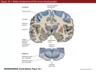
“The caudate in primates is part of the ‘associative striatum’, which receives inputs from association cortices. It corresponds to the dorsomedial striatum (caudate) in rodents, whereas the putamen is part of the sensorimotor striatum, corresponding to the dorsolateral striatum (putamen) in rodents.” 4. Use some diagrams to make clear the anatomy:
5. Discuss physical and functional differences between DLS and DMS systems. • Dorsal Striatum is made up of: • Caudate (medial) (upper left) • Putamen (lateral) (right) • Globus Pallidus (ventral) (lower left) • DLS & DMS differ in: • Connectivity • Distribution of various receptors • Mechanisms of synaptic plasticity
Dorsolateral Striatum (DLS) • Involved in stimulus-response learning • Controls movement and is connected to sensorimotor functioning (seeing, hearing, moving, etc.) • For example, when we move a part of our body in a new way and it feels good, for example salsa dancing, the dorsolateral striatum is active.
Dorsomedial Striatum (DMS) • Involved in action-outcome learning • Controls flexible behavior and is connected to areas where associations are recognized and formed • Belongs to the same functional system as the hippocampus. • For example, When we feel a sense of accomplishment for having tried something new, the association is recognized in the dorsomedial striatum.
6. Explain this statement: “It appears that because their habit system was disrupted by the lesion, the alternative A-O system assumed control over behavior . . . Because the DLS (the region thought to be involved in Stimulus-Response learning) had a lesion and was non-functional, the habit of lever pressing did not seem to be consolidated in these rats. After the value of the reward had been reduced and the rats were later tested for extinction, control rats pressed the levers at a higher rate than the rats with lesions to the DLS. It appears as if lever pressing had become a habit for the controls. It seems that when the Stimulus-Response type of learning cannot be performed by the DLS, the Action-Outcome type of learning compensates for this by becoming the primary type of learning to occur.
. . . However, a similar effect was not observed in rats with DMS lesions In this case, the DMS (the region thought to be involved in Action-Outcome learning) had a lesion and was non-functional. Contrary to the previous experiment, it appears that the Stimulus-Response type of learning is unable to compensate for the lack of Action-Outcome learning whenever the structures responsible for Action-Outcome learning are damaged.