rTMD123 Interaction with HMGB1 in Aorta and Inhibition of Binding to THP-1 Cells
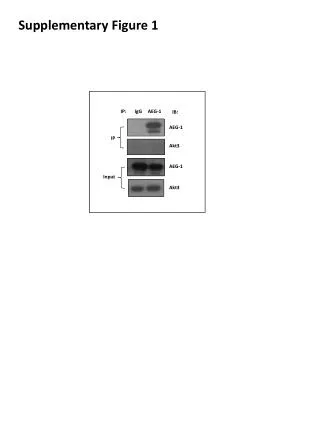
This study investigates the interaction between rTMD123, tagged with c-myc, and HMGB1 in aortic homogenates. Using rabbit anti-HMGB1 antibody and Protein A beads, we performed immunoprecipitation followed by western blot analysis to confirm the interaction. Additionally, we evaluated the inhibitory effect of rTMD123 on HMGB1 binding to THP-1 cells through flow cytometry (FACS), demonstrating a concentration-dependent response. The results reveal significant differences compared to untreated and HMGB1-treated groups, suggesting potential therapeutic implications.

rTMD123 Interaction with HMGB1 in Aorta and Inhibition of Binding to THP-1 Cells
E N D
Presentation Transcript
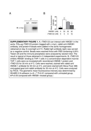
B A Mean fluorescence intensity 40 35 IgG HMGB1 30 IB: c-myc 25 HMGB1 (nM) 032 32 32 rTMD123 (nM) 0 0 8 16 SUPPLEMENTARY FIGURE 1. A, rTMD123 can interact with HMGB1 in the aorta. Fifty-μg rTMD123 protein (tagged with c-myc), rabbit anti-HMGB1 antibody, and protein A beads were added in the aortic homogenates (obtained on day 3) overnight at 4C. Rabbit IgG antibody (IgG) was served as a negative control. Beads were washed thrice with PBS containing 0.05% Tween-20 and the immune precipitates were analyzed by western blot. The result is typical of those obtained in 3 independent experiments. B, rTMD123 inhibits HMGB1 binding to THP-1 cells in a concentration-dependent manner. THP-1 cells were co-incubated with recombinant HMGB-1 protein and rTMD123 for 30 min at 4C. Cells were washed, stained with rabbit-anti HMGB-1 antibody for 30 min at 4C, and were stained with Alexa Fluor 488-conjugated goat anti-rabbit antibody for 30 min at 4C. Cells were analyzed by FACS. The geometric mean fluorescence intensity was determined by WinMDI 2.9 software (n=3). (**P<0.01 compared with untreated group. #P<0.05 compared with HMGB1-treated group.) IP