Download
1 / 1
10 likes | 92 Views
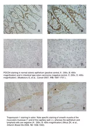
Examination of tropomyosin and PDCD4 staining in colon tissues, with specific staining observed in smooth muscle and capillary wall for tropomyosin, and contrasting staining in normal epithelium and colon carcinoma for PDCD4.

E N D
* * □ □ A B Tropomyosin 1 staining in colon. Note specific staining of smooth muscle of the muscularis mucosae (*) and of the capillary wall (□), whereas the epithelium and lymphoid cells are negative (A: 200x, B: 400x magnification) (Mirza ZK, et al., Inflamm Bowel Dis 2006, 12: 1036-1043). A B C D PDCD4 staining in normal colonic epithelium (positive control, A : 200x, B: 400x magnification) and in intestinal type colon carcinoma (negative control, C: 200x, D: 400x magnification). (Mudduluru G, et al., Cancer 2007, 110: 1697-1707.).