Download
1 / 29
300 likes | 634 Views
α -Synuclein occurs physiologically as a helically folded tetramer that resists aggregation. Tim Bartels, Joanna G. Choi, & Dennis J. Selkoe. Christopher O’Brien CHEM 645 Project Presentation 11/09/11. Protein misfolding and disease. Protein misfolding is the cause of a number of diseases

E N D
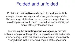
α-Synuclein occurs physiologically as a helically folded tetramer that resists aggregation Tim Bartels, Joanna G. Choi, & Dennis J. Selkoe Christopher O’Brien CHEM 645 Project Presentation 11/09/11
Protein misfolding and disease • Protein misfolding is the cause of a number of diseases • Neurodegenerative diseases are a well-studied subset • Misfolding of proteins is often the first step in an aggregation pathway • Formation of amyloid fibrils • α-synuclein is associated with the pathogenesis of Parkinson’s disease Dobson, C. M.(2001)
α-synuclein – role in disease • Implicated in molecular events leading to Parkinson’s disease • Lewy bodies characteristic of Parkinson’s disease contain aggregates of α-synuclein • Three point mutations are known to lead to familial forms of Parkinson’s disease: • A53T • A30P • E46K • Forms amyloid-like fibrils http://en.wikipedia.org/wiki/File:Lewy_Koerperchen.JPG T Ulmeret al. Journal of Biological Chemistry280, 9595-9603 (2005)
α-synuclein – natively unfolded protein? • Commonly believed to be natively unfolded monomer • Approximately 14 kDa • Crystal structure of micelle-bound α-synuclein shown to the left • Solved by NMR • Helical secondary structure only acquired with lipid binding • Protein obtained by recombinant bacterial overexpression
New methods for determination of native state of α-synuclein • Look at cell lines that endogenously express α-synuclein • M17D (dopaminergic human neuroblastoma) • HEK293 • HeLa • COS-7 • Other sources of native α-synuclein • Frontal cortex of wild-type mice • Human red blood cells • The above were examined using native gel electrophoresis
Native gel electrophoresis • Non-denaturing conditions (no SDS) • Proteins expected to remain folded in native state • Charged denaturing agent not used • Proteins may migrate in gel differently based on molecular mass, charge in gel conditions, as well as hydrodynamic size and shape • In this work, both BN-PAGE and CN-PAGE are used • CN-PAGE omits coomassie blue from sample preparation and cathode buffer
Native gel electrophoresis results • BN-PAGE shows an approximately 45-50 kDa molecular weight for all endogenous α-synuclein • CN-PAGE shows ~55-60 kDa, consistent with a tetramer • Monoclonal and polyclonal α-synuclein antibodies used for detection in both systems
SDS-PAGE results • In vitro crosslinking used to preserve assembled state • Tetramer bands observed in crosslinked SDS-PAGE
2D IEF/SDS-PAGE/Western blot analysis of crosslinked RBC lysate • Higher molecular weight oligomers have same isoelectric point as monomers
Purification of endogenous α-synuclein from living human cells • RBC in lysate were precipitated with (NH4)2SO4 • Three different purification methods • Hydrophobic interaction chromatography • Anion exchange chromatography • Covalent chromatography (activated thiopropyl sepharose) • Subsequent size exclusion step for further purification • Figure shows SDS-PAGE after three stages of purification
Scanning transmission electron microscopy (STEM) • Type of TEM • Electrons pass through thin sample • Electron beam focused onto single spot and scanned over sample • Tobacco mosaic virus (TMV) rods included during preparation as internal sizing standard • Measurements carried out at the Brookhaven National Laboratory
Sedimentation equilibrium analytical ultracentrifugation (SE-AUC) • Time-independent equilibrium concentration profile where sedimentation and diffusion are in equilibrium • Distributions are Boltzmann distributions • Insensitive to shape of molecules • Results fitted using software SEDPHAT to calculate molecular weight • Determined from average of three concentrations • Error and standard deviation calculated using Monte-Carlo simulations
Circular dichroism – looking at secondary structure • Measures the difference in adsorption of left and right polarized UV light • Ideal spectra for alpha helix, beta sheet, and random coil shown to the right • Spectra can be fit to multiple ideal spectra to determine content of different secondary structures in protein http://www.ap-lab.com/circular_dichroism.htm
CD spectra of RBC α-synuclein • Minima at 222 and 208 nm and overall shape indicative of α-helical secondary structure • Binding to lipid doesn’t appear to be required for folded structure • Random coil conformation seen in recombinant monomer expressed from bacteria
Analysis of CD spectra with Lipidex treatment • Lipidex 1000 = a reagent used to strip proteins of bound lipids and fatty acids • Used to confirm that lipid association is not required for helical structure
Quantitative elemental phosphate analysis • Used to verify lack of a significant phospholipid presence • Involves incubation with ammonium molybdate(VI) tetrahydrate and 10% ascorbic acid • Absorbance at 280nm measured and compared to calibration curve of 7 standards • Results indicated only 0.25mol phosphate per mole α-synuclein present • Significant presence of phospholipids on purified native α-synuclein unlikely
Examining post-translational modifications using mass spectrometry • MALDI-TOF used to analyze • (A) Recombinant αSyn • (B) Purified αSyn from human RBC • Predicted theoretical mass = 14,502 kDa • N-α-acetylation found on monomer from human RBC
Purification of stably transfected α-synuclein from human neuroblastoma cells • 3D5 cells, a M17D human neuroblastoma cell line was stably transfected to overexpress wild-type human α-synuclein • Endogenous α-synuclein from untransfected M17D cells also analyzed • Cells were lysed by sonication • Similar purification performed as described for RBCs using anion exchange chromatography
Comparison of stably transfected and RBC α-synuclein • Similar migration and CD spectra between both types of expression/purification
STEM measurements of 3D5 expressed α-synuclein • Very similar molecular weight to RBC α-synuclein
Comparison of α-synuclein from 3D5 and bacterial recombinant expression • Bacterial monomeric α-synuclein added extrinsically to parental M17D cell line prior to purification identical to 3D5 • Can conclude that α-helically folded α-synuclein doesn’t arise from artificial manipulation during lysis and purification
Surface plasmon resonance (SPR) • Used to measure adsorption of a material • Light beam used to excite surface plasmons (oscillating electrons in metal film) • Light incident at an angle greater than the critical angle is subject to total internal reflection • An evanescent wave interacts with surface plasmons and excites them • This is dependent on proteins bound to surface http://upload.wikimedia.org/wikipedia/commons/f/f8/Otto-schema.png
SPR results • RBC purified α-synuclein has a much greater resonance angle shift than recombinant monomer • Fit to two state model – dissociation constant two orders of magnitude lower than recombinant monomer
Thioflavin T (ThT) fluorescence • Used to quantify formation of amyloid fibril growth • Binds to beta sheet-rich structures and displays enhanced fluorescence with a red shift in its spectrum • Sample is incubated with ThT and fluorescence is measured from 460-550nm • Possibility of unwanted binding or spectroscopic change may cause unreliable results in quantitative analysis
ThT fluorescence results • Purified cellular α-synuclein incubated over 10 days shows no evidence of fibril formation sufficient for ThT binding • Recombinant monomer incubated for the same time shows amyloid fibril formation
Conclusions • Endogenous α-synuclein is natively an α-helical tetramer of about 58 kDa • This contradicts previously published work describing α-synuclein as a natively unfolded 14 kDa monomer • Variable amounts of lower number oligomers also present in cells • Based on the predominant native structure, it is likely that α-synuclein tetramers undergo destabilization before non-native aggregation and fibrillar assemblies that are characteristic of Parkinson’s disease form • Consistent with the observation that partial unfolding of a protein typically occurs before aggregation
Possible future directions • Further investigation into the native tetramer conformation of α-synuclein can be performed now that a protocol for its expression and purification is published • Closer examination of its mechanism of destabilization and aggregation • Therapeutic compounds that could kinetically stabilize native tetramers could also be investigated • Could lead to novel treatments for Parkinson’s and similar diseases