Cell Communication
Cell Communication. Evolution of Cell Signaling. A signal-transduction pathway is a series of steps by which a signal on a cell’s surface is converted into a specific cellular response Signal transduction pathways convert signals on a cell’s surface into cellular responses

Cell Communication
E N D
Presentation Transcript
Evolution of Cell Signaling • A signal-transduction pathway is a series of steps by which a signal on a cell’s surface is converted into a specific cellular response • Signal transduction pathways convert signals on a cell’s surface into cellular responses • Pathway similarities suggest that ancestral signaling molecules evolved in prokaryotes and have since been adopted by eukaryotes
Communication Between Mating Yeast Cells factor Exchange of mating factors. Each cell type secretes a mating factor that binds to receptors on the other cell type. Receptor a 2 3 1 factor Yeast cell, mating type a Yeast cell, mating type Mating. Binding of the factors to receptors induces changes in the cells that lead to their fusion. a New a/ cell. The nucleus of the fused cell includes all the genes from the a and cells. a/
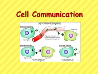
Cells Communication • Direct contact • Paracrine signaling • Endocrine signaling • Synaptic signaling
Direct Contact Plasma membranes Plasmodesmata between plant cells Gap junctions between animal cells Cell junctions Cell-cell recognition • Cells touch each other and signal molecules travel through special connections called communicating junctions • Communicating junctions link the cytoplasms of 2 cells together, permitting the controlled passage of small molecules or ions between them.
Cell Communication In Animals Local signaling Long-distance signaling Blood vessel Electrical signal along nerve cell triggers release of neurotransmitter Endocrine cell Target cell Neurotransmitter diffuses acrosssynapse Secretory vesicle Secreting cell Hormone travels in bloodstream to target cells Local regulator diffuses through extracellular fluid Target cell Target cell is stimulated (b) Synaptic signaling. A nerve cell releases neurotransmitter molecules into a synapse, stimulating the target cell. (a) Paracrine signaling. A secreting cell acts on nearby target cells by discharging molecules of a local regulator (a growth factor, for example) into the extracellular fluid. (c) Hormonal signaling. Specialized endocrine cells secrete hormones into body fluids, often the blood. Hormones may reach virtually all body cells. • In many other cases, animal cells communicate using local regulators, messenger molecules that travel only short distances • In long-distance signaling, plants and animals use chemicals called hormones
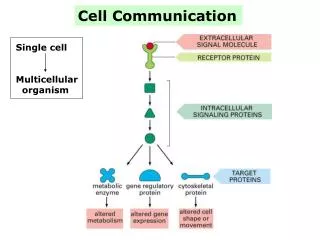
Cell Signaling EXTRACELLULAR FLUID CYTOPLASM Plasma membrane 3 1 2 Reception Transduction Response Receptor Activation of cellular response Relay molecules in a signal transduction pathway Signal molecule • The cells of a organism communicate with each other by releasing signal molecules that bind to receptor proteins located either on or inside of target cells. • Three stages of cell signaling: • Reception - each target cell has receptors that detect a specific signal molecule and binds to it • Transduction – binding of the signal molecule changes the receptor protein in some way that initiates transduction or conversion of the signal to a form that can bring about a specific cellular response • Response – transduced signal triggers a specific cellular response, any cell activity
Reception • A signal molecule binds to a receptor protein, causing it to change shape • The binding between signal molecule (ligand) and receptor is highly specific • A conformational change in a receptor • Is often the initial transduction of the signal
Receptors • Intracellular receptors • Some signal molecules that are small or hydrophobic can pass through the plasma membrane and bind to receptors located inside the cell • Intracellular receptors are cytoplasmic or nuclear proteins • Cell surface receptors. - Signal molecules that cannot pass through the plasma membrane bind to receptors located on the surface of the membrane
Intracellular Receptors • Gene Regulators • Signal molecule joins to the receptor, the receptor changes shape and a DNA binding site is exposed. • The DNA binding site joins to a specific segment of DNA and activates (or suppresses) a particular gene • Enzyme Receptor • These receptors function as enzymes – proteins that catalyze (speed up) specific chemical reactions. • When a signal molecule joins to the receptor, the receptor’s catalytic domain is activated (or deactivated).
Steroid hormone interacting with an intracellular receptor Hormone (testosterone) EXTRACELLULAR FLUID The steroid hormone testosterone passes through the plasma membrane. 4 3 2 5 1 Plasma membrane Testosterone binds to a receptor protein in the cytoplasm, activating it. Receptor protein Hormone- receptor complex The hormone- receptor complex enters the nucleus and binds to specific genes. DNA The bound protein stimulates the transcription of the gene into mRNA. mRNA NUCLEUS New protein The mRNA is translated into a specific protein. CYTOPLASM
Surface Receptors • Receptors located on the surface of the membrane, 4 types: • Chemically gated ion channels • Enzymatic receptors • G-protein-linked receptors • Integrins
Chemically Gated Ion Channels Gate close Gate Closed Signalmolecule(ligand) Ions Ligand-gated ion channel receptor Plasma Membrane Gate open Cellularresponse Gate close • An ion channel receptor acts as a gate when the receptor changes shape • When a signal molecule binds as a ligand to the receptor, the gate allows specific ions, such as Na+ or Ca2+, through a channel in the receptor
Enzymatic Receptors • Embedded in the plasma membrane, with their catalytic site exposed inside the cell. • Catalytic site activated when the signal molecule joins to the receptor. • Function as protein kinases (enzymes that phosphorylate proteins.)
Receptor Tyrosine Kinases Signal-binding site Signalmolecule Signal molecule Helix in the Membrane Tyr Tyr Tyr Tyr Tyrosines Tyr Tyr Tyr Tyr Tyr Tyr Tyr Tyr Receptor tyrosinekinase proteins(inactive monomers) Dimer CYTOPLASM Activatedrelay proteins Cellularresponse 1 P P Tyr Tyr Tyr Tyr P P Tyr Tyr Tyr Tyr P P P Tyr Tyr Tyr Tyr P Tyr Tyr Tyr Tyr Cellularresponse 2 P P P Tyr Tyr Tyr Tyr P Tyr Tyr Tyr Tyr 6 ATP 6 ADP Activated tyrosine- kinase regions (unphosphorylated dimer) Fully activated receptor tyrosine-kinase (phosphorylated dimer) Inactiverelay proteins
G-protein-linked Receptors Signal G-protein-linked receptor G protein Enzyme or ion channel Activated G protein Activated enzyme or ion channel • Signal molecule joins to a receptor, the receptor activates a G protein • The activated G protein can then activate an ion channel or enzyme in the plasma membrane.
Signal-binding site G-PROTEIN-LINKED RECEPTORS Segment that interacts with G proteins Inactive enzyme Activatedreceptor G-protein-linked receptor Signal molecule Plasma Membrane GDP G-protein(inactive) GTP GDP CYTOPLASM Enzyme Activated enzyme GTP GDP Pi Cellular response
Second Messengers • Some enzymatic receptors and most G-protein-linked receptors relay their message into the cell by activating other molecules or ions inside the cell. • These molecules and ions, called second messengers, transmit the message within the cell. The 2 most common second messengers are cAMP and Ca++
Signal Transduction Pathways • Transduction usually involves multiple steps • Multistep pathways • Can amplify a signal • Provide more opportunities for coordination and regulation • The molecules that relay a signal from receptor to response are mostly proteins • The receptor activates another protein, which activates another, and so on, until the protein producing the response is activated • At each step, the signal is transduced into a different form, usually a conformational change
A Phosphorylation Cascade Signal molecule Receptor Activated relay molecule A relay molecule activates protein kinase 1. 3 4 2 1 5 Active protein kinase 1 transfers a phosphate from ATP to an inactive molecule of protein kinase 2, thus activating this second kinase. Inactive protein kinase 1 Active protein kinase 1 Inactive protein kinase 2 Active protein kinase 2 then catalyzes the phos- phorylation (and activation) of protein kinase 3. ATP Phosphorylation cascade P Active protein kinase 2 ADP PP P i Inactive protein kinase 3 Enzymes called protein phosphatases (PP) catalyze the removal of the phosphate groups from the proteins, making them inactive and available for reuse. ATP P Finally, active protein kinase 3 phosphorylates a protein (pink) that brings about the cell’s response to the signal. ADP Active protein kinase 3 PP P i Inactive protein ATP P ADP Active protein Cellular response PP P i • In many pathways, the signal is transmitted by a cascade of protein phosphorylations - phosphatase enzymes remove the phosphates • This phosphorylation and dephosphorylation system acts as a molecular switch, turning activities on and off
cAMP Second MessengerG-protein-signaling pathway First messenger (signal molecule such as epinephrine) Adenylyl cyclase G protein GTP G-protein-linked receptor ATP Second messenger cAMP Protein kinase A Cellular responses • Signal molecule binds to surface receptor • Surface receptor activates a G protein • G protein activates the membrane-bound enzyme, adenylyl cyclase • Adenylyl cyclase catalyzes synthesis of camp, which binds to a target protein • Target protein initiates cellular change
Cyclic AMP NH2 NH2 NH2 N N N N N N N N N N N O O O N O Adenylyl cyclase Phoshodiesterase CH2 O HO Ch2 P –O O P O P P O CH2 O O O O O O O O O P Pyrophosphate H2O O O P P i OH OH OH OH OH ATP Cyclic AMP AMP • Cyclic AMP (cAMP) is one of the most widely used second messengers • Adenylyl cyclase, an enzyme in the plasma membrane, converts ATP to cAMP in response to an extracellular signal
Calcium (Ca++) Pathways EXTRACELLULAR FLUID Plasma membrane Ca2+ pump ATP Mitochondrion Nucleus CYTOSOL Ca2+ pump Endoplasmic reticulum (ER) ATP Ca2+ pump Low [Ca2+] High [Ca2+] Key • Calcium ions (Ca2+) act as a second messenger in many pathways • Calcium is an important second messenger because cells can regulate its concentration
Calcium ions and Inositol Triphosphate (IP3) • A signal transduction pathway may trigger an increase in calcium in the cytosol • Pathways leading to the release of calcium involve inositol triphosphate (IP3) and diacylglycerol (DAG) as second messengers
Ca++ Pathway • Signal molecule binds to surface receptor • Surface receptor activates a G protein • G protein activates the membrane-bound enzyme, phospholipase C • Phospholipase C catalyzes synthesis of inositol triphosphate, which stimulates release of Ca++ from ER • Released Ca++ initiates cellular change
Calcium and IP3 in signaling pathways 2 4 3 5 1 6 A signal molecule binds to a receptor, leading to activation of phospholipase C. DAG functions as a second messenger in other pathways. Phospholipase C cleaves a plasma membrane phospholipid called PIP2 into DAG and IP3. EXTRA- CELLULAR FLUID Signal molecule (first messenger) G protein DAG GTP PIP2 G-protein-linked receptor Phospholipase C IP3 IP3-gated calcium channel Endoplasmic reticulum (ER) Various proteins activated Cellularresponses Ca2+ Ca2+ (second messenger) CYTOSOL The calcium ions activate the next protein in one or more signaling pathways. IP3 quickly diffuses through the cytosol and binds to an IP3– gated calcium channel in the ER membrane, causing it to open. Calcium ions flow out of the ER (down their con- centration gradient), raising the Ca2+ level in the cytosol.
Fine-Tuning of the Response • Multistep pathways have two important benefits: • Amplifying the signal (and thus the response) • Contributing to the specificity of the response • Enzyme cascades amplify the cell’s response • At each step, the number of activated products is much greater than in the preceding step
Amplification • Due to the many steps in the cell signaling process, one signal molecule can trigger a “cascade” effect
Cytoplasmic response to a signal: the stimulation of glycogen breakdown by epinephrine Reception Binding of epinephrine to G-protein-linked receptor (1 molecule) Transduction Inactive G protein Active G protein (102 molecules) Inactive adenylyl cyclase Active adenylyl cyclase (102) ATP Cyclic AMP (104) Inactive protein kinase A Active protein kinase A (104) Inactive phosphorylase kinase Active phosphorylase kinase (105) Inactive glycogen phosphorylase Active glycogen phosphorylase (106) Response Glycogen Glucose-1-phosphate(108 molecules)
Specificity of Cell Signaling Signal molecule Receptor Relay molecules Response 2 Response 3 Response 1 Cell A. Pathway leads to a single response Cell B. Pathway branches, leading to two responses Activation or inhibition Response 5 Response 4 Cell D. Different receptor leads to a different response Cell C. Cross-talk occurs between two pathways • Different kinds of cells have different collections of proteins • These differences in proteins give each kind of cell specificity in detecting and responding to signals • The response of a cell to a signal depends on the cell’s particular collection of proteins • Pathway branching and “cross-talk” further help the cell coordinate incoming signals
Signaling Efficiency: Scaffolding Proteins and Signaling Complexes Signalmolecule Plasmamembrane Receptor Threedifferentproteinkinases Scaffoldingprotein Figure 11.16 • Rather than relying on diffusion of large relay molecules such as proteins, many signal pathways are linked together physically by scaffolding proteins. • Scaffolding proteins may themselves be relay proteins to which several other relay proteins attach. • This hardwiring enhances the speed, accuracy, and efficiency of signal transfer between cells.