Cell Communication: Understanding the Cellular Internet
Explore how multicellular organisms communicate to maintain balance through cell signaling methods using chemical messengers and cell interactions. Discover the importance of local and long-distance signaling with examples and the three stages of cell signaling pathway. Learn about receptors and response activation for effective cellular communication.

Cell Communication: Understanding the Cellular Internet
E N D
Presentation Transcript
The “Cellular Internet” • All multicellular organisms must “communicate and cooperate” to maintain homeostasis • Science has identified universal (meaning ALL life) mechanisms of cell-to-cell communication. • Communication in cells is similar to communication in general. Cells communicate by sending/receiving signals and then converting the signals into a response
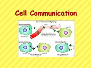
Communication Methods • Cell Signaling occurs using chemical messengers (proteins, steroids, electrical impulses, etc) that pass from one cell to another • Local signalingoccurs over short distances • Ex: Cell-Cell Recognition Proteins attached to cell exterior; glycolipids and glycoproteins (e.g. blood type proteins); cells must TOUCH at least temporarily • Ex: Local regulators (chem signals from the neighboring cells); communication between cells in the same area • Long distance signaling occurs over larger distances • Ex: Hormones (chemical messengers in the form of proteins or steroids) are put into the blood and travel through the body until they reach the target cell
Plasma membranes Plasmodesmata between plant cells Gap junctions between animal cells Figure 11.3 (a) Cell junctions. Both animals and plants have cell junctions that allow molecules to pass readily between adjacent cells without crossing plasma membranes. LOCAL SIGNALING EXAMPLES • Transport between cells • cell junctions are protein tunnels directly connecting adjacent cells (called gap junctions in animal cells & plasmodesmata in plants); allow material to pass through (e.g. chem signals or water) and be shared
(b) Cell-cell recognition. Two cells in an animal may communicate by interaction between molecules protruding from their surfaces. Figure 11.3 LOCAL SIGNALING EXAMPLES • Cell to Cell Recognition: Cell “ID badges” (glycoproteins and glycolipids) are used to identify cells that belong; cells without the proper identification are attacked as invaders; part of immune response • “ID badges” are membrane bound cell surface molecules that are checked by membrane bound receptor proteins on the immune system cells
Local signaling (Left: Paracrine, Right: Synaptic) Target cell Electrical signal along nerve cell triggers release of neurotransmitter Neurotransmitter diffuses acrosssynapse Secretory vesicle Local regulator diffuses through extracellular fluid Target cell is stimulated LOCAL SIGNALING EXAMPLES - Local Regulators: Chemical signals used to communicate with neighboring cells within a tissue, only work over a short distance - Paracrine signaling communicates with all cells surrounding and coordinates efforts (e.g. growth factors are released to stimulate mitosis in all cells near a wound to promote healing) - Synaptic signaling occurs when the signal is directed to only one neighbor cell (e.g. neurotransmitters pass from one neuron to the next to send a signal to the brain)
Long-distance signaling Blood vessel Endocrine cell Hormone travels in bloodstream to target cells Target cell (c) Hormonal signaling. Specialized endocrine cells secrete hormones into body fluids, often the blood. Hormones may reach virtually all body cells. LONG DISTANCE SIGNALING EXAMPLES • used by all multicellular organisms (plant and animal) to coordinate effort between cells that are not close together; hormone (natural steroids or proteins) chemical signals released directly into vascular tissue or even the air by glands can go anywhere in the body or even to another organism; target cells contain surface receptors that recognize and respond to the hormone while other cells ignore the signal; slow method of communication, but it can cause changes in a lot of cells simultaneously (e.g. adrenal response)
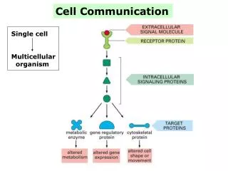
Three Stages of Cell Signaling • All cell signaling (long or short distance) occurs in three stages • Reception – cell receives the signal (called a ligand) at the membrane • Transduction – signal causes a cascade of communication inside the cell to relay the message from the membrane to the appropriate interior organelle • Response – cell responds to the signal by generating an appropriate change • Called Signal transduction pathways • Note: Pathways may vary in the specifics, but all living things use these three steps; support for evolution
EXTRACELLULAR FLUID CYTOPLASM Plasma membrane 1 2 3 Reception Transduction Response Receptor Activation of cellular response Relay molecules in a signal transduction pathway Signal molecule Figure 11.5 Overview of cell signaling
Stage One: Reception • The signaling molecule (called a ligand, but could be any hormone/protein/ion/etc) binds to a specific receptor protein; shape determines function! Only cells that contain the right receptor can receive the signal CYTOPLASM EXTRACELLULAR FLUID Plasma membrane Reception 1 1 The receptor and signaling molecules fit together (lock and key model, induced fit model, just like enzymes!) Receptor Signaling molecule
Receptor Proteins Examples G-protein coupled receptors Receptor tyrosine-kinases Ion channel receptors
G-Protein Coupled Receptors are often involved in diseases such as bacterial infections. G-Protein Receptors Inactive enzyme Plasma membrane G protein-coupled receptor Activated receptor Signaling molecule Enzyme GDP 2 1 GDP GTP CYTOPLASM G protein (inactive) Activated enzyme i GTP GDP P 4 3 Cellular response
Signal-binding site Signalmolecule Signal molecule Helix in the Membrane Tyr Tyr Tyr Tyr Tyrosines Tyr Tyr Tyr Tyr Tyr Tyr Tyr Tyr Receptor tyrosinekinase proteins(inactive monomers) Dimer CYTOPLASM Activatedrelay proteins Cellularresponse 1 Tyr Tyr Tyr Tyr Tyr Tyr P P Tyr P Tyr Tyr Tyr Tyr P Tyr Tyr Tyr P P P Tyr Tyr Tyr Tyr Tyr P Tyr Tyr Tyr Cellularresponse 2 P P P Tyr Tyr P 6 ATP 6 ADP Activated tyrosine- kinase regions (unphosphorylated dimer) Fully activated receptor tyrosine-kinase (phosphorylated dimer) Inactiverelay proteins Figure 11.7 Receptor tyrosine kinases
Ion Channel Receptors Gate closed 1 Ions Signaling molecule (ligand) • Used by the nervous system • Signal triggers the opening of an ion channel which allows a rush of ions into the cell, creating a burst of electrical activity called an action potential • Must be used in conjunction with a concentration gradient (dam metaphor) and reset each time Ligand-gated ion channel receptor Plasma membrane 2 Gate open Cellular response 3 Gate closed
Stage Two: Transduction CYTOPLASM EXTRACELLULAR FLUID Plasma membrane Reception Transduction 1 1 2 Receptor 2nd Messenger! Relay molecules in a signal transduction pathway Signaling molecule • Reception sets off a “relay team” of INTERIOR communication molecules; proteins and/or second messengers (non-proteins) carry the original exterior signal to the inside of the cell
Notes about Transduction • It is a multistep pathway • can amplify (increase) a signal and create a large response from a single ligand • Requires communication and coordination within the cell itself
Transduction Example • Protein Phosyphorylation Cascade - An example of transduction in which a series of proteins called kinases add a phosphate to the next one in line, activating it, and sending the signal to the target (like a bucket brigade!) • enzymes then remove the phosphates to reset the cascade
Signal molecule A relay molecule activates protein kinase 1. Receptor Activated relay molecule 4 1 3 5 2 Inactive protein kinase 1 Active protein kinase 1 transfers a phosphate from ATP to an inactive molecule of protein kinase 2, thus activating this second kinase. Active protein kinase 1 Active protein kinase 2 then catalyzes the phos- phorylation (and activation) of protein kinase 3. Inactive protein kinase 2 ATP Phosphorylation cascade P Active protein kinase 2 ADP PP P i Enzymes called protein phosphatases (PP) catalyze the removal of the phosphate groups from the proteins, making them inactive and available for reuse. Inactive protein kinase 3 Finally, active protein kinase 3 phosphorylates a protein (pink) that brings about the cell’s response to the signal. ATP P ADP Active protein kinase 3 PP P i Inactive protein ATP P ADP Active protein Cellular response PP P i • A phosphorylation cascade Figure 11.8
Transduction Example • Second Messengers • Secondary messengers are small, NON-PROTEIN molecules or ions that carry the signal to the target organelle; (Note: Membrane Proteins would be the primary messengers since they get the signal first)
First messenger (signal molecule such as epinephrine) Adenylyl cyclase G protein GTP G-protein-linked receptor ATP cAMP Protein kinase A Cellular responses Secondary Messenger Example: Cyclic AMP • cAMP is made from ATP but has only one phosphate and is reattached to itself (circular shape); acts as a secondary messenger Figure 11.10
NH2 NH2 NH2 N N N N N N N N N N N O O O N O Adenylyl cyclase Phoshodiesterase CH2 O HO Ch2 P –O O P O P P O CH2 O O O O O O O O O P Pyrophosphate H2O O O P P i OH OH OH OH OH ATP Cyclic AMP AMP Cyclic AMP • Cyclic AMP (cAMP) • made from ATP
First messenger Adenylyl cyclase G protein Fig. 11-11 GTP G protein-coupled receptor ATP Second messenger cAMP Ex Diagram: Transduction in a G-protein pathway using cAMP Protein kinase A Cellular responses
EXTRACELLULAR FLUID Plasma membrane Ca2+pump ATP Mitochondrion Nucleus CYTOSOL Ca2+pump Endoplasmic reticulum (ER) ATP Ca2+pump Key High [Ca2+] Low [Ca2+] Second Messenger Example: Calcium Ions • Calcium ions also act as a secondary messenger because cells can regulate the concentration and location (dam metaphor again!) Other secondary messengers trigger the release of concentration gradients of Ca2+ in various areas of the cell, creating moving charges and electrical signals. Pumps then reset the Ca2+ concentration gradient to be used again.
6 3 2 1 4 5 A signal molecule binds to a receptor, leading to activation of phospholipase C. DAG functions as a second messenger in other pathways. Phospholipase C cleaves a plasma membrane phospholipid called PIP2 into DAG and IP3. EXTRA- CELLULAR FLUID Signal molecule (first messenger) G protein DAG GTP PIP2 G-protein-linked receptor Phospholipase C IP3 (second messenger) IP3-gated calcium channel Endoplasmic reticulum (ER) Various proteins activated Cellularresponse Ca2+ Ca2+ (second messenger) The calcium ions activate the next protein in one or more signaling pathways. IP3 quickly diffuses through the cytosol and binds to an IP3– gated calcium channel in the ER membrane, causing it to open. Calcium ions flow out of the ER (down their con- centration gradient), raising the Ca2+ level in the cytosol. Calcium Ion Diagram example Figure 11.12
Stage Three: Response CYTOPLASM EXTRACELLULAR FLUID Plasma membrane Reception Transduction Response 1 2 3 Receptor Activation of cellular response Relay molecules in a signal transduction pathway Can be catalysis, activation of a gene, triggering apoptosis, almost anything! Signaling molecule • The cell will respond to the signal as directed (e.g. make a protein, produce more energy, enter mitosis, etc.)
Signaling molecule • Specificity of the signal • The same signal can trigger different responses depending on the receiving cell; example – adrenaline increases the activity of heart cells but slows digestive cells • Many responses can come from one signal! Receptor Relay molecules Response 1 Response 2 Response 3 Cell A. Pathway leads to a single response. Cell B. Pathway branches, leading to two responses.
Cell responses vary widely; signals can activate, inhibit ,or create multiple responses Activation or inhibition Response 4 Response 5 Cell C. Cross-talk occurs between two pathways. Cell D. Different receptor leads to a different response.
Response example- cell signaling can lead to regulation of transcription (turn genes on to make a needed protein or off to stop production)
Response Example Hormones often induce transcription. Once inside the cell, the hormone attaches to a protein that takes it into the nucleus where transcription can be stimulated. (ex: testosterone, which is a transcription factor that stimulates muscle and hair growth)
Termination of Communication • Response is terminated quickly by the reversal of ligand binding; once the signal is degraded or released, the response will conclude
Any Questions?? Can You Hear Me Now?
Organ System Communication Specialists • Nervous System (Animals only) • Quick long distance communication done through chains of local synaptic signals that pass electrical stimulation through the cells themselves and chemicals called neurotransmitters in between neighboring cells; FAST, but limited in scope • Endocrine System (Animals only) • Glands (lymph nodes, adrenal gland, pituitary gland, etc)that secrete hormones into cell spaces or the blood stream to coordinate cells throughout the body; SLOW, but can generate a large response (ex: menstruation, fight or flight) • Note: Plants also use hormones, but each cell releases hormones individually (no gland type tissues) • Plant hormomes are transported through vascular system, plasmodesmata, or released into air (e.g. ripening fruit)
Major Vertebrate Endocrine Glands Their Hormones (Hypothalamus–Parathyroid glands)
Neurosecretory cells in endocrine organs and tissues secrete hormones. These hormones are excreted into the circulatory system (ex. Adrenaline) or the surrounding cell space (ex. Lymph).
Are the following hormone pathways Positive or Negative Feedback systems? Stress and the Adrenal Gland http://highered.mcgraw-hill.com/olcweb/cgi/pluginpop.cgi?it=swf::535::535::/sites/dl/free/0072437316/120109/bio48.swf::Action%20of%20Epinephrine%20on%20a%20Liver%20Cell
http://bcs.whfreeman.com/thelifewire/content/chp42/4202003.htmlhttp://bcs.whfreeman.com/thelifewire/content/chp42/4202003.html
http://vcell.ndsu.nodak.edu/animations/regulatedsecretion/movie.htmhttp://vcell.ndsu.nodak.edu/animations/regulatedsecretion/movie.htm
Answers • Stress and Adrenaline – Positive Feedback (induces a response/change) • Calcium and Blood Sugar regulation – Negative Feedback (prevents a change, maintains a normal level)