Download
1 / 72
720 likes | 1.57k Views
Endotracheal Intubation. Types of endotracheal tubesPlain Used in birdsOccasionally used in catsA plain tube does not form an airtight seal within the tracheaWill allow secretions, blood or gastro-intestinal fluid to enter the lungs. Endotracheal Intubation. Types of endotracheal tubesCuffed Used in all speciesCuff is inflated with air to produce a leak proof sealA pilot balloon is used to estimate degree of inflationAlternatively the cuff is filled until no leak of air can be heard wh1144

E N D
1. Intubation and Anesthetic Machines
2. Endotracheal Intubation Types of endotracheal tubes
Plain
Used in birds
Occasionally used in cats
A plain tube does not form an airtight seal within the trachea
Will allow secretions, blood or gastro-intestinal fluid to enter the lungs
3. Endotracheal Intubation Types of endotracheal tubes
Cuffed
Used in all species
Cuff is inflated with air to produce a leak proof seal
A pilot balloon is used to estimate degree of inflation
Alternatively the cuff is filled until no leak of air can be heard when the lungs are inflated to 25 cm H2O pressure
4. Endotracheal Intubation Types of endotracheal tubes
Cole
Originally designed for babies
The tube has excellent pressure-flow characteristics and provides less resistance to breathing
Absence of cuff allows insertion of a larger tube
A cole tube is commonly used for cats
"Shoulders" of the tube form an airtight seal at the entrance to the larynx
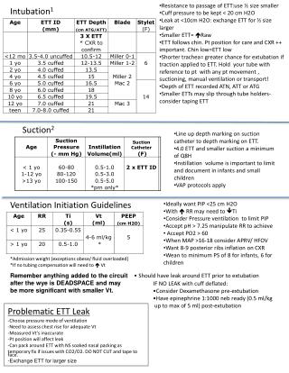
5. Endotracheal Intubation Size of endotracheal tube
Choose an endotracheal tube as large as possible without forming a "push-fit�
Check the tube length alongside the animal
The tip of the tube must be beyond the larynx but not extend past the thoracic inlet
6. Endotracheal Intubation Size of endotracheal tube
Tube sizes refer to the internal diameter of the part of the tube residing in the trachea
A few manufacturers print both internal and external diameters on their endotracheal tubes
Purchase tubes with thin walls especially when they are to be used on cats and small dogs
A thin wall allows a tube with a larger lumen to be used
7. Endotracheal Intubation Technique of intubation
Orotracheal Intubation
Dogs and Cats
Open the dog or cat's mouth and view the larynx
Use a bright overhead light or a laryngoscope
There is no excuse for doubt when inserting an endotracheal tube
You can see where it is -- in the trachea or in the esophagus
8. Endotracheal Intubation Technique of intubation
Orotracheal Intubation
Dogs and Cats
Pull the tongue forward gently in the dog and cat to move the larynx more rostral . Do not pull hard on the tongue or it will could lacerated or have nerve damage
Do not put your fingers inside the mouth (jaw tone and the ability to reflexively close the jaws persists into light anesthesia)
A plastic or metal stilette can be used inside an endotracheal tube to stiffen it
This is to improve control of the direction of the tip of the tube, not so that you can force the tube into the larynx
Make sure that the tip of the stilette is not sharp and does not extend beyond the end of the tube, where it might lacerate the trachea
9. Endotracheal Intubation Technique of intubation
Orotracheal Intubation
Dogs and Cats
If you use a laryngoscope
Place the tip of the blade over the tongue and under the epiglottis
Placing it on the epiglottis will distort the laryngeal opening and make it harder to intubate
Tracheal intubation can be performed with the animal on its side, sternum, or back
The position may be dictated by the animal's condition (e.g., with GI obstruction always intubate the animal in sternal position, with the head up) or by personal preference of the surgeon
12. Endotracheal Intubation Technique of intubation
Orotracheal Intubation
Pigs and small ruminants
Technique used is the same for dogs and cats
The anatomy of the pharynx and larynx of these species differs
Adult Cattle
Intubation can be performed using a laryngoscope with an extra long blade
Intubation most frequently accomplished without viewing the larynx
You introduce your hand and arm into the mouth to palpate the laryngeal opening and to guide the tip of the endotracheal tube into the trachea
Horses
Intubation performed "blind", without viewing the larynx and without palpation.
13. Nasotracheal Intubation Dogs, cats, pigs and ruminants
Not performed
Horses
Easy to insert a tube from the nares through the ventral nasal meatus, into the trachea
Used in some foals to administer halothane or isoflurane to induce anesthesia
Used in adult horses during recovery from anesthesia to relieve nasal obstruction from mucosal swelling
14. Pharyngostomy Intubation Tracheal intubation through a pharyngostomy
Dogs and cats
May be used in dogs and cats requiring oral surgery, e.g., repair of fractured jaw, cleft palate repair, lacerations of hard palate
Allows the surgeon greater access to the surgical field
15. Exotics Reptiles � Most reptiles can be intubated if large enough.
Avian � Endotracheal intubation sometimes used. High incidence of mucous blockage in tubes due to very small tracheal openings and high mucous production. Most avian veterinarians prefer air sac cannulization. This is a surgical procedure when a tracheal tube is inserted directly into an air sac near the leg.
16. Endotracheal Intubation Complications of endotracheal intubation
If the internal diameter of the endotracheal tube is too small in relation to the animal
There will be increased resistance to breathing
Hypoventilation will result
If the cuff is over inflated, or if it is inflated for too long
The tracheal mucosa, and sometimes the cartilage, will die and slough
Later the animal will show signs of tracheitis
Only inflate the tube cuff with enough air to prevent a leak when the animal's lungs are artificially inflated
If you have a device to measure intra-cuff pressure, inflate the cuff to 25 cm H2O pressure
If the duration of surgery is long, provided that regurgitation has not occurred, the cuff should be deflated and the tube repositioned every 2 hours
17. Endotracheal Intubation Complications of endotracheal intubation
If the tube is too long
A primary bronchus may be intubated
One lung will collapse
Cyanosis may develop
Delivery of inhalation anesthetic is impaired
The animal may wake up
Endotracheal tubes can cause airway obstruction
They become twisted or kinked
The bevel of the tube lies against the tracheal wall
The cuff is over inflated and squashes the tube lumen
The cuff bulges over the end of the tube
K-Y lubricant is allowed to dry inside the tube
Mucus and secretions accumulate during anesthesia (especially cats and small dogs) or are allowed to dry inside the tube (improper cleaning after anesthesia).
18. Endotracheal Intubation Complications of endotracheal intubation
Trauma of the larynx and trachea
Can produce laryngitis/tracheitis which will last for several days
Laryngeal trauma may also predispose the animal to laryngospasm or to granuloma formation at a later date
Mucosal swelling in the recovery period may cause partial or complete airway obstruction
Preferably do not cause trauma, but if swelling occurs, inject dexamethasone IV and provide supportive treatment (oxygen, reintubate) until the swelling is reduced
Irregular heart rhythm
Laryngoscopy causes bradycardia
Tracheal intubation causes tachycardia and, sometimes, ventricular premature depolarizations
Atropine premedication should prevent bradycardia
Lidocaine, 2 mg/kg, applied directly to the larynx or injected intravenously before intubation should modify or prevent tachycardia
Lidocaine is used when tachycardia is anticipated to cause deterioration of the patient.
19. Endotracheal Intubation Complications of endotracheal intubation
Leak in the cuff preventing an airtight seal and controlled ventilation
Always check the cuff for leaks before inducing anesthesia
Transfer of infection
Clean tubes thoroughly, inside and outside, between patients
Use a bristle brush and betadine (or similar) solution
If ethylene oxide (ETO) sterilization is used, adequate aeration is needed to eliminate irritant residues
20. Endotracheal intubation Complications of endotracheal intubation
Bracycephalic syndrome (short nose breeds)
Stenotic nares
Everted layrngeal saccules
Elongated soft palate
Hypoplastic trachea
Airways can collapse easily after extubation
Wait until the dog has a good gag reflex before extubating
Preoxygenate for 5 minutes before induction in case of a difficult intubation
21. Endotracheal Intubation Complications of endotracheal intubation
Cats
Can be very difficult to intubate due to laryngeal spasms
Small amount of lidocaine swabbed on larynx can numb it long enough to intubate
Many need to use a stylet in endotracheal tube to help pass it
22. Endotracheal Intubation Complications of endotracheal intubation
Collapsing trachea
Genetically weak trachea
Preoxygenate severe cases
Intubate as gently as possible as not to irritate the trachea
23. Endotracheal Intubation Procedure for tracheal extubation in dogs and cats
Leave tube in place until swallowing reflex returns
Remove blood clots from nasopharynx
Deflate the cuff before extubation
Exceptions:
When bleeding into the nose and mouth has occurred
Excessive salivation has occurred
Regurgitation has occurred
Prevent the animal biting the tube while it is being removed
Cost prohibitive
Animal (dog or cat) can easily inhale or swallow pieces of tube
24. Parts of an Anesthetic Machine Gas cylinders
Oxygen and nitrous oxide are contained in compressed gas cylinders
Found as E cylinders that are usually attached to the machine via yokes that are equipped with a specific pin system
Tanks also come in large G and H cylinders
25. Parts of an Anesthetic Machine Gas cylinders
Pressure gauge attached to the cylinder indicates the pressure of the gas in the tank
Pressure in a full O2 cylinder is 2200 psi
Oxygen tanks should be changed when pressure drops below 500 to 600 psi
The volume of O2 in an E cylinder can be calculated by multiplying the psi by 0.3
A full tank of 2200 psi will contain 660 L of O2
26. Parts of an Anesthetic Machine Gas cylinders
Nitrous tanks are stored at lower pressures
A full tank is 770 psi
Nitrous tanks should be changed when pressure gauge drops below 500 psi
Both liquid and gas states are present but the gauge reads only the gas state
Liquid evaporates to gas as soon as the gas leaves the tank so the pressure in the tank will not change until all the liquid state has evaporated
28. Parts of an Anesthetic Machine Pressure releasing valve (regulator)
Reduces the high pressure of the O2 or nitrous leaving the tank to a low pressure of 50 psi
29. Parts of an Anesthetic Machine Flow meter
Measure O2 or nitrous in L/min
Allows the anesthetist to set the O2 or nitrous oxide flow rates that will be delivered to the animal
As the gas passes through the flow meter gas pressure is reduced further to 15 psi
31. Parts of an Anesthetic Machine Vaporizer
Converts the liquid anesthetic into a gas state
Controls the amount of vaporized amount of vaporized anesthetic mixed with the carrier gas
33. Parts of an Anesthetic Machine Check valves
Inhalation/Exhalation flutter valves
Insures a uni-directional flow of gas to and from the patient when delivering a circle system
Y connector
Connects the endotracheal tube to the inspiratory and expiratory tubes of a circle system
34. Parts of an Anesthetic Machine Rebreathing bag (reservoir bag)
Allows the animal to breath easier from a reservoir of gas
Can be used to deliver O2 (with or without anesthetic gas) and manually assist respirations, bagging
Bags should have a minimum volume of 60 ml/kg of patient weight
35. Parts of an Anesthetic Machine Carbon dioxide absorber
Soda lime canister
Used in rebreathing systems to remove CO2 from the expired gases
Exhaust gases enter a canister containing soda lime or barium hydroxide
Na+, K+, Ca2+ and Ba2+ hydroxide reacts with the exhaled CO2 and water to form carbonate
Heat is liberated and the pH decreases
36. Parts of an Anesthetic Machine Carbon dioxide absorber
Soda lime canister
A pH color indicator turns blue on consumption
When the soda lime or barium hydroxide granules turn color or the granules become hard instead of crumbly, they are saturated with CO2 and should be replaced
When in use, the granules will produce heat and condensation within the canister
The color reaction is time limited
Exhausted crystals should be removed immediately and replaced with new granules
37. Parts of an Anesthetic Machine Carbon dioxide absorber
Soda lime canister
Should be changed after 6 to 8 hours of use depending on the size of the animal and the gas flow rate
If machines are left standing for longer than 30 days, granules should be replaced before using machine
38. Parts of an Anesthetic Machine Exhaust valve
Also called the pop-off valve, or pressure relief valve
Exhaust gases leave the system via the exhaust valve entering the scavenger system
Valve can be fully or partially open when a patient is using the machine
Valve is closed for leak tests or when filling the reservoir for assisted respirations
39. Parts of an Anesthetic Machine Manometer
Measures the pressure in the system in mm Hg or cm H20
Generally calibrated form -30 to +50 cm H20
Gauge thus reflects the pressure of gas in the animal�s airways and lungs
The pressure should be at 0 and never more than 15 cm H20 (11 mm Hg)
When providing positive assisted ventilation, the pressure should not exceed 15 to 20 cm H20 (11 to 15 mm Hg)
40. Parts of an Anesthetic Machine Oxygen flush valve
O2 bypasses vaporizer, delivering 100% O2 to breathing system
Enables the anesthetist to flush the system with pure O2
Fills the reservoir and system for leak test
Also flushes the anesthetic gases out of the circuit and replaces with pure oxygen
Never use O2 flush valve with a Bain circuit in a small animal because it produces too much pressure
41. Parts of an Anesthetic Machine Scavenger system
Attached to the exhaust valve
Consists of tubing that collects gases and directs them outside the building or to a charcoal canister
Can be active or passive
42. Parts of an Anesthetic Machine Negative pressure relief valve
Some newer machines have this safety feature
Valve opens in response to a negative pressure situation in the system
Allows room air into the circuit
Negative pressure could be due to an active scavenger system or a low oxygen supply
43. Maintenance Oxygen tanks must be turned off to prevent excess pressure on the regulator
Flush the remaining O2 to minimize damage to the pressure gauge and reducing valves
Turn flowmeter off to prevent sudden rush of O2 into the flowmeter when O2 is turned back on
Don�t over tighten because the knobs can be easily twisted off
44. Maintenance After each anesthesia induction, removable machine parts and anesthetic equipment that come in contact should be washed in a mild, soapy solution, soaked in a cold disinfectant, thoroughly rinsed and dried
The dome valves and absorbent canister should be disassembled and wiped dry.
Flutter valves need periodic removal and cleaning with a disinfectant to prevent adherence to the machine housing
45. Maintenance Vaporizers should be turned off when not in use and periodically emptied to prevent buildup of the preservative and other residue
Best to clean and recalibrate by authorized personnel every 6 to 12 months
Isoflurane does not contain a preservative
46. Maintenance Barium hydroxide or soda lime granules found in the CO2 absorbers need replacing when the granules have changed color or cannot be easily crumbled
Do not tightly pack and leave about 1 cm (1/2 inch) of air space
Avoid having dust enter tubing or hoses of the machine
Rubber items will likely need to be replaced after prolonged use
47. Environmental concerns Environmental pollution can be minimized through proper equipment use and scavenging of the gases
Safe exposure limit for inhalant anesthetic agents has been set at 2 p.p.m. in room air
Everyone, especially pregnant women, should avoid high levels of waste anesthetic gases
Much of the anesthetic levels are because of leaks in anesthetic machines
48. Environmental concerns Vaporizers and CO2 absorbers should be filled with minimal personnel in a well ventilated area while wearing gloves and masks
Do not turn the vaporizer on and off until or while, the patient is connected to the machine
During recovery, keep patient in a well ventilated area and on the machine until expired gases are scavenged
49. Environmental concerns Use active scavenging systems whenever possible to ensure waste gases are drawn out of the area
If passive scavenging is used, keep the hose as short as possible and have it travel downward toward the exhaust
If it is not possible to install scavengers in all rooms where machines are used, either use an activated charcoal cartridge that must be replaced after 12 hours or substitute injectable anesthesia
50. Environmental concerns Before anesthesia, the machine should be checked for both high and low pressure leaks
Leakage of nitrous is the major environmental concern
A high pressure system test monitors NO2 and O2 leakage
A low pressure system leak is in the anesthetic machine itself
51. Environmental concerns Low pressure system test
A low pressure system leak occurs between the flowmeter and the patient
Turn the tank on, close the pop-off valve, and occlude the end of the hose so the gas should have nowhere to escape
Adjust the flowmeter to at least 2 L/min of O2 allowing the bag to fill gradually and then turn off the flowmeter
If there is no escape of air when the bag is gently squeezed, then there is no low pressure system leakage
52. Environmental concerns Low pressure system test
System can also be checked by occluding as above and using the flowmeter to allow the system to pressure at 30 cm H2O
Turn off the flowmeter
The pressure should be maintained for at least 10 seconds
One can also listen for the hiss of escaping air or use a detergent solution as described earlier
53. Breathing Systems Rebreathing systems
Circle systems
Rebreathing refers to breathing a mixture of expired gases and fresh gases
The amount of CO2 in inhaled gases depends on
Whether the rebreathing system has a CO2 absorber
The flow rate of fresh gases (the higher the fresh gas flow rate, the more expired gas is pushed out the scavenger and not rebreathed
Depending on the flow rate of fresh gas, the system is classified as a closed system (total rebreathing of expired gases) or semi-closed system (partial rebreathing of expired gases)
54. Breathing Systems Closed rebreathing systems
With closed systems the fresh gas flow rate is does not exceed the patient�s metabolic O2 consumption of 5 to 10 mL/kg/min
The system may be used with a closed pop-off valve and a fresh gas flow rate of 5 to 10 mL/kg/min
Expired gases are recirculated (after CO2 removal) with incoming fresh gases
55. Breathing Systems Closed rebreathing systems
Danger of increased CO2 accumulation if CO2 absorber not working efficiently
It is economical and there is minimal pollution
It takes longer to change planes of anesthesia
O2 depletion and N2O buildup are common, so do not use N2O with this system
Requires constant monitoring to ensure pressures do not build up in the system if the O2 flow delivered exceeds the metabolic requirement
56. Breathing Systems Closed rebreathing systems
It can be dangerous to run a rebreathing system with the pop-off valve closed, if the pop-off valve does not have a safety release at high pressures
It is recommended that the pop-off valve be left partially open to prevent increases in pressure in the system and to adjust the O2 flow rate accordingly to prevent the rebreathing bag from collapsing
If the bag does not collapse you can be confident that sufficient O2 is being delivered to meet the patients metabolic requirements
57. Breathing Systems Semi-closed or partial rebreathing systems
With semi-closed systems, the fresh gas is delivered in excess of metabolic consumption at 25 to 50 mL/kg/min (suggested economical flow rate)
The gas escapes through the pop-off valve to the scavenger or after having the CO2 removed by the soda lime and then recirculated with the fresh gases
Higher flow rates can be used
Less rebreathing will occur
N2O buildup is less of a concern with higher flow rates
Important to flush the system to prevent nitrogen buildup from the expired gases
58. Breathing Systems Non-rebreathing systems
There is no mixing of inhaled and exhaled gases and no rebreathing of expired gases; all expired gas goes to the scavenger
CO2 absorber not required
Fresh gas flow rates required at 200 to 300 mL/kg/min
Fresh gas flow rates required at 130 to 200 mL/kg/min with the Bain system
May be some rebreathing of exhaled gases if a reservoir bag and low flow rate
59. Breathing Circuits Many kinds of breathing circuits available
Circle system
Universal F-circuit
Bain system (Coaxial)
60. Breathing Circuits Circle system
CO2 absorber
Inspiratory and expiratory unidirectional valves (check valves or flutter valves)
Two breathing hoses connected with a Y-piece to the patient
Rebreathing bag
Pop-off valve (exhaust valve)
scavenger
61. Breathing Circuits Circle system
Can be used as a non-rebreathing system (200 mL/kg/min)
Can be used as a partial rebreathing system (25 to 50 mL/kg/min)
Can be used as a total rebreathing system (5 to 10 mL/kg/min)
62. Breathing Circuits Circle system
An advantage is the mixture of expired gases with incoming gases
Humidifies and warms the incoming gases
Main disadvantages of the circle system occur with smaller patients
Excess weight and bulk of the hoses
Excess dead space
Resistance to breathing caused by the unidirectional valves
63. Breathing Circuits Universal F-circuit
Basically a modified circle system where the inspiratory hose is placed within the expiratory hose
Still requires a CO2 absorber, rebreathing bag, unidirectional valves, pop-off valve and scavenger
64. Breathing Circuits Universal F-circuit
Incoming fresh gas is warmed also by expired gases
The advantage is lighter weight and less bulk
Disadvantage is that if the system is stretched, the end of the inspiratory hose pulls away from the end of the expiratory hose
Considered a safety feature so that the hoses don�t break
Increases the dead space within the circuit
Even when not stretched, the dead space is equivalent to the circle system
65. Breathing Circuits Bain system
Consists of one tube inside the other
Fresh gases flow through the inner tube
Unused and exhaled gases flow through the outer tube
There also is a rebreathing bag with a clip on the tubing between the reservoir bag and scavenger connection but no CO2 absorber
Between breaths, the fresh gases flow through the inner tube toward the patient and then back through the outer tube toward the scavenger
66. Breathing Circuits Bain system
When the patient inspires, the gases are drawn from the inner tube, which will be 100% fresh gases or a mixture of fresh gases and expired gases, depending on the fresh gas flow rate
This system can be used as a non-rebreathing system with a fresh gas flow rate of 200 to 300 mL/kg/min
The high flow rate pushes exhaled gases away down the outer tube so there is no rebreathing of exhaled gases
By changing the flow rate to 130 to 200 mL/kg/min the system acts as a partial rebreathing system
Most of the gases get pushed away but there is partial rebreathing of some exhaled gases
67. Breathing Circuits Bain system
Ideal for small patients (<7kg)
Lightweight
Minimal dead space
Little resistance to breathing
Good for all small animals in general but is not economical when the patient weighs in excess of 10kg
Limiting factor is the size of the patient
The O2 flowmeter must provide flow rates required for a partial or non-rebreathing system (130 to 300 mL/kg/min)
Total volume of the Bain hose must be greater than the tidal volume of respiration of the patient to effectively prevent rebreathing
68. Breathing Circuits Bain system
Good for procedures involving the head (less tubing in the way)
Good for procedures with much manipulation (i. e. radiography, because there is less weight pulling on the head)
Warming and humidification are minimal with partial rebreathing
Requires a precision vaporizer
69. Vaporizers Vapor pressure is characterized by the amount of vapor related to its liquid in a closed container
The pressure exerted by the gas is called the vapor pressure and will increase with increases in temperature
Most anesthetics vaporize at a concentration higher than necessary for clinical anesthesia
So a vaporizer is used to deliver diluted anesthetics to patients
70. Vaporizers Precision vaporizer
Enables delivery of controlled concentrations of anesthetic vapor independent of time, temperature and fresh gas flow rate
Temperature and flow rate are compensated for by the vaporizer or manually by the anesthetic technician
71. Vaporizers VOC (vaporizer out of circle)
Vaporizer is added to the system between the O2 flowmeter and the circle
The circle consists of the inspiratory and expiratory valves, breathing hoses,CO2 absorber, pop-off valve, scavenger and rebreathing bag
72. Vaporizers VIC ( vaporizer in circle)
Vaporizer is placed inside the breathing system, usually between the inspiratory valve and the patient
VIC�s are always non-precision
The carrier gas passes over the surface of the anesthetic liquid or past a wick
Incoming gases mix with warm exhaled gases in the system
Better vaporization of liquid is obtained when low fresh gas flows are used
High flows cool liquid and reduce vaporization
Are also safest when used with agents with low vapor pressure (e. g. methoxyflurane)