Download
1 / 58
620 likes | 1.32k Views
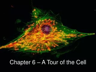
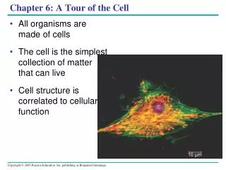
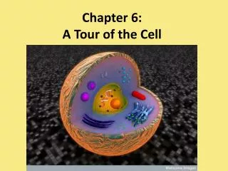
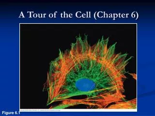
A Tour of the Cell (Chapter 6). Figure 6.1. Theodore Schwann’s cell theory (1839):. 1) All organisms are composed of one or more cells 2) Cells are the basic units of organization of all organisms 3) Cells arise only by division of a previously existing cell. How do we study cells?.

E N D
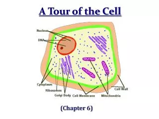
A Tour of the Cell (Chapter 6) Figure 6.1
Theodore Schwann’s cell theory (1839): • 1) All organisms are composed of one or more cells • 2) Cells are the basic units of organization of all organisms • 3) Cells arise only by division of a previously existing cell
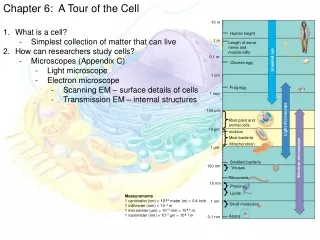
How do we study cells? Few cells are big enough to be viewed with the naked eye. Therefore, we use: • Cytology (microscopy) – study of cell structure - magnification – ratio of object’s image size to its real size - resolution – measure of clarity of the image, minimum distance two points can be separated and still be distinguished as two points - limits the effective study
Microscopy • Light microscopes (LMs) • First used during the Renaissance (1600s) • Pass visible light through a specimen • Magnify cellular structures with lenses
10 m Human height 1 m Length of some nerve and muscle cells 0.1 m Light microscope Chicken egg 1 cm Frog egg 1 mm 100 µm Most plant and Animal cells Electron microscope 10 µ m NucleusMost bacteriaMitochondrion 1 µ m Electron microscope Smallest bacteria 100 nm Viruses 10 nm Ribosomes Proteins Lipids 1 nm Small molecules Figure 6.2 Atoms 0.1 nm Smaller subcellular structures require higher powered microscopes 1 micrometer = 1 micron = 0.000001 or 1/100,000 meter = 1 nanometer = 0.000000001 or 1/100,000,000 meter =
RESULT TECHNIQUE (a) Brightfield (unstained specimen). Passes light directly through specimen. Unless cell is naturally pigmented or artificially stained, image has little contrast. [Parts (a)–(d) show a human cheek epithelial cell.] 50 µm (b) Brightfield (stained specimen).Staining with various dyes enhances contrast, but most staining procedures require that cells be fixed (preserved). (c) Phase-contrast. Enhances contrast in unstained cells by amplifying variations in density within specimen; especially useful for examining living, unpigmented cells. Figure 6.3 Microscopy • Different methods for enhancing visualization of cellular structures
(d) (e) Fluorescence. Shows the locations of specific molecules in the cell by tagging the molecules with fluorescent dyes or antibodies. These fluorescent substances absorb ultraviolet radiation and emit visible light, as shown here in a cell from an artery. 50 µm (f) Confocal. Uses lasers and special optics for “optical sectioning” of fluorescently-stained specimens. Only a single plane of focus is illuminated; out-of-focus fluorescence above and below the plane is subtracted by a computer. A sharp image results, as seen in stained nervous tissue (top), where nerve cells are green, support cells are red, and regions of overlap are yellow. A standard fluorescence micrograph (bottom) of this relatively thick tissue is blurry. 50 µm Research Methods Differential-interference-contrast (Nomarski). Like phase-contrast microscopy, it uses optical modifications to exaggerate differences in density, making the image appear almost 3D.
TECHNIQUE RESULTS 1 µm Cilia Scanning electron micro- scopy (SEM). Micrographs taken with a scanning electron micro- scope show a 3D image of the surface of a specimen. This SEM shows the surface of a cell from a rabbit trachea (windpipe) covered with motile organelles called cilia. Beating of the cilia helps move inhaled debris upward toward the throat. (a) • Electron microscopes (EMs) • Focus a beam of electrons through a specimen (TEM) or onto its surface (SEM) • The scanning electron microscope (SEM) • Provides for detailed study of the surface of a specimen
TEM The transmission electron microscope (TEM) • Provides for detailed study of the internal ultrastructure of cells Longitudinal section of cilium Cross section of cilium 1 µm (b) Transmission electron micro- scopy (TEM). A transmission electron microscope profiles a thin section of a specimen. Here we see a section through a tracheal cell, revealing its ultrastructure. In preparing the TEM, some cilia were cut along their lengths, creating longitudinal sections, while other cilia were cut straight across, creating cross sections. Figure 6.4 (b)
Electron micrographs of rabbit trachea cells Transmission EM Scanning EM Figure 6.4 - Beam through object - beam on surface
How do we study cells? 1) Microscopy 2) Cell fractionation - allows for determination of the function of subcellular structures • Take cells apart and separate the major organelles from each other • Done in a centrifuge – spin tubes at various speeds • Isolates cell components, based on size and density • Ultracentrifuges – most powerful, can spin as fast as 130K/minute = applies forces on particles of more than 1 million times the force of gravity
Homogenization Tissue cells 1000 g (1000 times the force of gravity) 10 min Homogenate Differential centrifugation Supernatant poured into next tube 20,000 g 20 min 80,000 g 60 min Pellet rich in nuclei and cellular debris 150,000 g 3 hr Pellet rich in mitochondria (and chloro- plasts if cells are from a plant) Pellet rich in “microsomes” (pieces of plasma mem- branes and cells’ internal membranes) Pellet rich in ribosomes Cell Fractionation Cells are homogenized in a blender to break them up. The resulting mixture (cell homogenate) is then centrifuged at various speeds and durations to fractionate the cell components Increasing speed (force) Results: 1)Researchers used microscopy to identify the organelles in each pellet 2) Researchers used biochemical methods to determine the metabolic functions of organelles. Big small Objects in the pellet Figure 6.5
Cells • Two types of cells • Prokaryotic • Eukaryotic • All cells have several basic features in common • Bounded by a plasma membrane • Contain a semifluid substance - cytosol • Contain chromosomes • Have ribosomes
Two types of cells • Circular DNA molecule (chromosome), not contained within a nucleus • Have their DNA located in a region called the nucleoid • Small in size 1-10 micrometers • Prokaryotic cells – basic, No interior membranes 2) Eukaryotic cells - Interior membranes • Linear DNA molecules (chromosomes) Contained within a true nucleus, bounded by a membranous nuclear envelope • 10-100 micrometers • Have extensive and elaborately arranged internal membranes, which form organelles
Prokaryotic cells Pili: attachment structures on the surface of some prokaryotes Nucleoid: region where the cell’s DNA is located (not enclosed by a membrane) Ribosomes: organelles that synthesize proteins Plasma membrane: membrane enclosing the cytoplasm Cell wall: rigid structure outside the plasma membrane Capsule: jelly-like outer coating of many prokaryotes Bacterialchromosome 0.5 µm Flagella: locomotion organelles of some bacteria (a) A typical rod-shaped bacterium (b) A thin section through the bacterium Bacillus coagulans (TEM) Figure 6.6
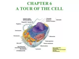
Animal cell (eukaryotic) Nuclear envelope ENDOPLASMIC RETICULUM (ER) NUCLEUS Nucleolus Smooth ER Rough ER Chromatin Flagelium Plasma membrane Centrosome CYTOSKELETON Microfilaments Intermediate filaments Ribosomes Microtubules Microvilli Golgi apparatus Peroxisome In animal cells but not plant cells: Lysosomes Centrioles Flagella (in some plant sperm) Lysosome Mitochondrion Figure 6.9
Plant Cell (eukaryotic) Nuclear envelope Rough endoplasmic reticulum Nucleolus NUCLEUS Chromatin Smooth endoplasmic reticulum Centrosome Ribosomes (small brown dots) Central vacuole Tonoplast Golgi apparatus Microfilaments Intermediate filaments CYTOSKELETON Microtubules In plant cells (but not in Animal cells): Chloroplasts Central vacuole and tonoplast Cell wall Plasmodesmata Mitochondrion Peroxisome Plasma membrane Chloroplast Cell wall Plasmodesmata Figure 6.9 Wall of adjacent cell
Prokaryotes vs. Eukaryotes Prokaryotes Eukaryotes Size 1-10 mm 10-100 mm Genetic circular linear Material chromosome chromosomes Plasma yes yes Membrane Cell Wall yes some-plants, etc. Ribosomes yes yes Membrane-bound NOYES organelles
Why are cells so small? • 1) Diffusion • main way of intracellular communication • diffusion is very slow! • 2) Surface area-to-volume ratio • A smaller cell - has a higher surface to volume ratio, which facilitates the exchange of materials into and out of the cell • volume increases more rapidly than surface area • Cell must receive nutrients and expel wastes across the surface area of the cell • Large volume = greater nutrient need and more waste production • consider a cube
Surface area and volume Surface area increases while total volume remains constant 5 1 1 Total surface area (height width number of sides number of boxes) 6 150 750 Total volume (height width length number of boxes) 125 125 1 Surface-to-volume ratio (surface area volume) 6 12 6 Area is proportional to the surface area squared Volume is proportional to the surface area cubed
TEM of a plasma membrane. The plasma membrane, here in a red blood cell, appears as a pair of dark bands separated by a light band. (a) Plasma Membrane • functions as a selective barrier • Allows sufficient passage of nutrients and waste Outside of cell Carbohydrate side chain Hydrophilic region Inside of cell 0.1 µm Hydrophobic region Hydrophilic region Phospholipid Proteins (b) Structure of the plasma membrane Figure 6.8 A, B
Nucleus • Contains the eukaryotic cell’s genetic instructions • Chromosomes, structures that carry genetic information • Made of chromatin (a complex of protein and DNA) • Each typical human cells has 46 chromosomes, 23 pairs • Fruit flies – 8 chromosomes • Dog – 78 • Cat - 38 • Enclosed by the nuclear envelope (double membrane), separating its contents from the cytoplasm
Nucleus-genetic information center Nucleus Nucleus 1 µm Nucleolus Chromatin Nuclear envelope: Inner membrane Outer membrane Nuclear pore Pore complex Rough ER Surface of nuclear envelope. 1 µm Ribosome 0.25 µm Close-up of nuclear envelope Nuclear lamina (TEM). Pore complexes (TEM).
Nuclear envelope • Nucleus is surrounded by a double membrane • Transport to-and-from cytoplasm via nuclear pores Inner membrane – lined with nuclear lamina Nuclear Pore Complex nucleus cytoplasm ER Outer membrane
Nucleolus • Densely stained portion of the nucleus • Site of ribosomal RNA (rRNA) synthesis • Site of beginning of ribosomal subunit assembly
Ribosomes - protein-producing machinery Ribosomes Cytosol Endoplasmic reticulum (ER) Free ribosomes Bound ribosomes Large subunit Bound Ribosomes - make proteins for insertion into membranes, packaging within certain organelles, or export from the cell Free Ribosomes – produce proteins which function in cytosol Small subunit 0.5 µm TEM showing ER and ribosomes Diagram of a ribosome
Ribosomessite of protein synthesis Large subunit -composed of protein and ribosomal RNA (rRNA) Small subunit - assembled in the nucleolus - active in the cytoplasm - prokaryotes have a smaller version (70S) than eukaryotes (80S) - actively growing or secreting eukaryotic cell may have millions of ribosomes
Endomembrane system • Extensive network of membranes • Accounts for more than half of the total membrane in many eukaryotic cells • Regulates protein synthesis and traffic • Performs metabolic functions in the cell • Metabolism and movement of lipids • The endomembrane system • Includes many different structures – nuclear envelope, endoplasmic reticulum, Golgi apparatus, lysosomes, vacuoles
The endoplasmic reticulum (ER) membrane • Is continuous with the nuclear envelope Smooth ER Nuclear envelope Rough ER ER lumen Cisternae Ribosomes Transitional ER Transport vesicle 200 µm Smooth ER Rough ER
Endoplasmic reticulum (ER)Accounts for more than half the total membrane in many eukaryotic cells 1) Smooth ER - no/few ribosomes - lipid synthesis (example -testes and ovaries) - carbohydrate metabolism - drug detoxification (liver cells) - calcium ion storage 2) Rough ER - ribosomes attached - continuous with nuclear membrane - Produces proteins and membranes, which are distributed by transport vesicles (pancreatic cells) - protein glycosylation (sugars added)
Golgi apparatus • Consists of flattened membranous sacs called cisternae • Polarity – opposite sides of the stack differ in thickness and molecular composition, cis face (receiving) and trans face (shipping) • Functions • Modification of the products of the rough ER • Manufacture of certain macromolecules • Receives many of the transport vesicles produced in the rough ER • Products of ER are modified (glycosylation), stored, sent out • Vesicles pinch off and carry cargo to appropriate locations in the cell
5 6 3 4 1 2 Golgi Apparatus functions cis face (“receiving” side of Golgi apparatus) Vesicles coalesce to form new cis Golgi cisternae Vesicles move from ER to Golgi 0.1 0 µm Vesicles also transport certain proteins back to ER Cisternae Cisternal maturation: Golgi cisternae move in a cis- to-trans direction Vesicles form and leave Golgi, carrying specific proteins to other locations or to the plasma mem- brane for secretion Vesicles transport specific proteins backward to newer Golgi cisternae trans face (“shipping” side of Golgi apparatus) Figure 6.13
Lysosome – acidic digestive compartments 1 µm Nucleus • Is a membranous sac of hydrolytic enzymes (acidic environments) from ER to Golgi to lysosome • Lysosomes carry out intracellular digestion by • Phagocytosis Lysosome Hydrolytic enzymes digest food particles Food vacuole fuses with lysosome Lysosome contains active hydrolytic enzymes Digestive enzymes Lysosome Plasma membrane Digestion Food vacuole (a) Phagocytosis: lysosome digesting food
Lysosome-acidic digestive compartment Lysosome containing two damaged organelles 1 µ m Mitochondrion fragment Peroxisome fragment Lysosome fuses with vesicle containing damaged organelle Hydrolytic enzymes digest organelle components Lysosome Digestion Vesicle containing damaged mitochondrion (b) Autophagy: lysosome breaking down damaged organelle • Autophagy – • Process of lysosomes which use their hydrolytic enzymes to recycle the cell’s own organic material • Damaged organelle or small amount of cytosol • Cell renews itself • Tay-Sachs disease • Lipid accumulation Figure 6.14 B
Central vacuole Cytosol Tonoplast Nucleus Central vacuole Cell wall Chloroplast 5 µm Vacuoles: Diverse Maintenance Compartments • A plant or fungal cell • May have one or several vacuoles • Food vacuoles • Are formed by phagocytosis • Contractile vacuoles • Pump excess water out of protist cells • Central vacuoles • Are found in plant cells • Hold reserves of important organic compounds and water • Surrounded by tonoplast
The endomembrane system • Complex and dynamic • Relationships among organelles of the endomembrane system 1 Nuclear envelope is connected to rough ER, which is also continuous with smooth ER Nucleus Rough ER 2 Membranes and proteins produced by the ER flow in the form of transport vesicles to the Golgi Smooth ER cis Golgi Nuclear envelop 3 Golgi pinches off transport Vesicles and other vesicles that give rise to lysosomes and Vacuoles Plasma membrane trans Golgi 4 5 6 Lysosome available for fusion with another vesicle for digestion Transport vesicle carries proteins to plasma membrane for secretion Plasma membrane expands by fusion of vesicles; proteins are secreted from cell
Cell Power! • Mitochondria and chloroplasts change energy from one form to another • Enclosed by membranes • not part of the endomembrane system • Mitochondria • cellular respiration • Chloroplasts • photosynthesis
Mitochondrion Intermembrane space Outer membrane Free ribosomes in the mitochondrial matrix Inner membrane Cristae Matrix Mitochondrial DNA 100 µm Mitochondria: Chemical Energy Conversion • Mitochondria are enclosed by two membranes • Smooth outer membrane • Inner membrane folded into cristae • Matrix – contains enzymes, • mitochondrial • DNA, and • ribosomes • divide and • partition • into daughter • cells Figure 6.17
Chloroplasts: Capture of Light Energy • Chloroplast - Contains chlorophyll • Specialized member of a family of plant organelles called plastids • Found in leaves/other green organs of plants and algae Chloroplast Stroma the internal fluid Ribosomes Chloroplast DNA Inner and outer membranes Granum 1 µm Thylakoid - membranous sacs
Peroxisomes • Removal of electrons and hydrogen - oxidation • Produce hydrogen peroxide as a byproduct, by transferring hydrogen from substrates to oxygen • Hydrogen peroxide is broken down to water • Break down fatty acids, detoxify alcohol/toxins • highly reactive and very destructive to cell • Contain catalase 2 H2O2 2 H2O + O2 catalase
Chloroplast Peroxisome Mitochondrion 1 µm Peroxisomes: Oxidation Often have a granular or crystal core - thought to be a dense collection of enzymes molecules
Microtubule Microfilaments 0.25 µm Figure 6.20 Cytoskeleton: Support, Motility, and Regulation • Network of fibers - organizes structures and activities in the cell • Gives mechanical support to the cell
Vesicle ATP Receptor for motor protein Motor protein (ATP powered) Microtubule of cytoskeleton (a) Motor proteins that attach to receptors on organelles can “walk” the organelles along microtubules or, in some cases, microfilaments. Vesicles Microtubule 0.25 µm (b) Vesicles containing neurotransmitters migrate to the tips of nerve cell axons via the mechanism in (a). In this SEM of a squid giant axon, two vesicles can be seen moving along a microtubule. (A separate part of the experiment provided the evidence that they were in fact moving.) Figure 6.21 A, B Cytoskeleton: Support, Motility, and Regulation • Involved in cell motility, which utilizes motor proteins • Railroad tracks for the cell, Trafficking of vesicles and organelles
Cytoskeleton Three main types of fibers make up the cytoskeleton
Cytoskeleton Table 6.1
Microtubules • Microtubules • Shape the cell • Guide movement of organelles • Help separate the chromosome copies in dividing cells • The centrosome • Is considered to be a “microtubule-organizing center”
Centrosome Microtubule Centrioles 0.25 µm Longitudinal section of one centriole Cross section of the other centriole Microtubules Figure 6.22 Centrosomes and Centrioles
(b) Motion of cilia. Cilia have a back- and-forth motion that moves the cell in a direction perpendicular to the axis of the cilium. A dense nap of cilia, beating at a rate of about 40 to 60 strokes a second, covers this Colpidium, a freshwater protozoan (SEM). (a) Motion of flagella. A flagellum usually undulates, its snakelike motion driving a cell in the same direction as the axis of the flagellum. Propulsion of a human sperm cell is an example of flagellatelocomotion (LM). Direction of swimming Figure 6.23 1 µm Cilia and Flagella • Specialized arrangements of microtubules • Are locomotor appendages of some cells Direction of organism’s movement Direction of Active stroke Direction of Recovery stroke
Cilia and flagella share a common ultrastructure Cilia – shorter than flagella - more abundant/cell than flagella • Cilia – shorter than flagella • more abundant/ • cell than flagella Outer microtubule doublet Plasma membrane 0.1 µm Dynein arms Central microtubule Outer doublets cross-linking proteins inside Microtubules Radial spoke Plasma membrane (b) Cross-section through the Cilium shows “9+2” arrangement Of microtubules Basal body 0.5 µm 0.1 µm (a) A longitudinal section of cilium shows microtubules running the length of the structure Triplet Figure 6.24 A-C (c) Basel Body – (anchor) nine outer doublets of a cilium or flagellum exend into the basel body where each doublet joins another microtubule to form a ring of nine triplets Cross section of basal body
Muscle cell Actin filament Myosin filament Myosin arm Myosin motors in muscle cell contraction. (a) Figure 6.27 A Microfilaments (Actin Filaments) • Found in microvilli and muscle cells • For cellular motility - Contain the protein myosin in addition to actin Microvillus Plasma membrane Microfilaments (actin filaments) Intermediate filaments 0.25 µm Figure 6.26