Download
1 / 49
510 likes | 552 Views
Understand how to calculate your mid-term grades based on three grades received so far. Learn methods for effective studying to achieve desired grades in courses. Includes helpful study tips and suggested techniques.

E N D
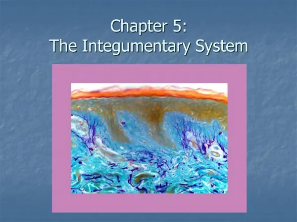
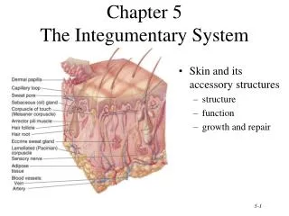
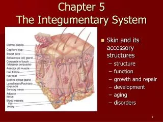
Visual Anatomy & PhysiologyFirst EditionMartini & Ober Chapter 5 Integumentary System Lecture 12
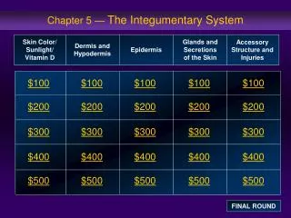
Mid-term Grades (based on 3 grades) - Revised Based on the three (3) grades you have received so far, you should do a mid-term checkup to see how you’re doing. To find your average so far, total the three grades you’ve received and divide by 300 (the total amount of points possible so far for the courses). Ex: keeping all grades: (83 + 50 + 90) 300 = 0.74 (74%) Ex: dropping the low grade: (83 + 90) 200 = 0.86 (86%) To figure out what you need to AVERAGE on the next lecture and two lab exams plus the final COMBINED to get your desired grade for the course : Pts desired (from syllabus) - Total pts. so far Average grade needed on remaining exams = 450 (if no grade dropped) or 550 (if low grade dropped)
Points and Grades (from Syllabus) - Revised Example 1: To get a grade of B for the course, using the example grades on previous slide, and not dropping lowest grade (50), and assuming 50 pts for lab and 4 XC points: 656 – (83 + 50 + 90 + 50 + 4) = x; x = 0.84 (84%) Average on upcoming exams 450 Example 2: To get a grade of B for the course, using the example grades on previous slide, and dropping lowest grade (50), and assuming 50 pts for lab and 4 XC points: 656 – (83 + 90 + 50 + 4) = x; x = 0.78 (78%) Average on upcoming exams 550
Suggested Study Method • 1. Read the lecture material (see my suggestions below) before coming to class and TAKE NOTES from the material. Here’s where the expert questions (the sheet I gave you on the first day) come in handy: as you read ask yourself these questions about the particular topic you’re reading, e.g, what’s it made of, where is it found, what makes it different from other things, how does it work, what if it doesn’t work correctly, etc. Remember to answer the expert questions for BOTH anatomy and physiology, where applicable. • 2. Take some notes in class if you need to, and then soon after class COMBINE you reading notes for that topic with your class notes. • 3. After you’ve had some time away from the material, CONDENSE your notes for that topic as much as you can. • 4. After you’ve condensed, then do a preliminary study of your notes, using various things (mnemonics) to help you remember them. • 5. Once you’ve geared up to start studying for a test, try and CONDENSE your notes a little more without sacrificing clarity and review them and re-memorize anything you’ve forgotten. REMEMBER, when studying for an exam, you are now using ONLY your notes, not your textbook (unless you need clarification about something). Make sure you’ve gone over your notes and memorized then AT LEAST three times before the exam • 6. Finally, get a BLANK Study Guide, put your notes aside then answer the questions on the study guide and compare your answers to your notes. If you can’t answer the questions correctly or address the points on your Study Guide without looking at your notes, you’re not yet ready for the exam. Review/re-memorize those things you are still not solid on. • A suggested method for reading your text is the following: • 1. Skim the chapter section HEADINGS first to get an overview of the chapter contents • 2. Look at all the figures in the text and read the legends for the figures/tables before you actually read. Ask yourself what you know about the figures as you’re looking at them. • 3. Read over the Study Guide for the material (that I give you for each exam) before you read to focus your thoughts on the most important 20% of what you will have to know. • 4. Read the Chapter summary (yes, BEFORE you read the Chapter) and while you’re reading through it keep asking yourself what you already know and what you don’t know, and focus on the things that are mentioned in your Study Guide. • 5. Read the chapter material and, using the Study Guide and the Expert Questions, make notes on what you’re reading. Don’t COPY sentences in your textbook word-for-word, but put a summary sentence for each paragraph you read IN YOUR OWN WORDS.
Lecture Overview • Functions of the Integumentary System • Overview of the skin • The epidermis • The dermis • The hypodermis (subcutaneous layer) • Accessory structures of the integumentary system • Injury and Repair • Aging and the integumentary system
Some Questions… Skin is composed of an epithelial layer and a connective tissue layer. Is skin a membrane? Yes. A cutaneous membrane What is a membrane? Combination of ET and CT tissues combine to protect/cover other tissues Is there a difference between the skin (integument) and the integumentary system? Yes. Skin is the cutaneous membrane consisting of an epithelium (epidermis) and a dermis (CT). The integumentary system (IS) includes the skin, hair, nails, and glands (accessory structures). How does the structure of the the integument, enable it to perform its functions of protection, temperature regulation, etc.? That is the subject of this lecture…
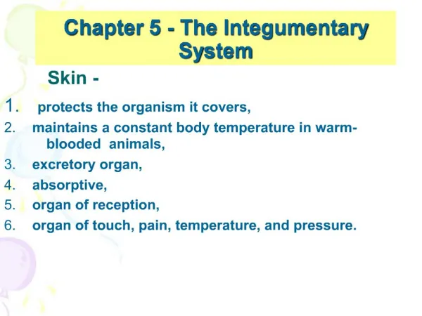
Introduction to the Integumentary System • The integument constitutes 16% of our body weight and has a surface area of about 1.5 – 2.0 m2 (15 – 20 ft2) • Functions of the integument • Protection (from mechanical/chemical/bacterial damage, UV radiation) • Temperature regulation (extreme heat, extreme cold) and Fluid conservation • Excretion • Vitamin D production • Sensation (touch, pressure)
Overview of the Integument SKIN Accessory Structures Epidermis = protection; Dermis = nourishment of epidermis; SubQ = insulation Figure from: Martini, Anatomy & Physiology, Prentice Hall, 2001
Cells of the Epidermis • Epidermis of the skin is classified as a keratinized stratified squamous epithelium • Cells of the epidermis include • Keratinocytes (90%) • Keratin – a tough, fibrous intracellular protein (protection) • Lamellar granules (waterproofing, extracellular) • Melanocytes (8%) • Produce melanin (protection from UV radiation) • Langerhans cells(1-2%) • Migrate to skin from bone marrow • Participate in skin’s immune response (dendritic cells) • Merkel cells(< 1%) • Least numerous; specialized epithelial cells • Function in sensation of touch
Thick and Thin Skin Thin (0.07-0.12 mm)(epidermal thickness) Thick (0.8-1.4 mm)(epidermal thickness) Thick skin - palms of hands, soles of feet; fiveepidermal layers Thin skin - everywhere else; fourepidermal layers (no s. lucidum) Figure from: Martini, Anatomy & Physiology, Prentice Hall, 2001
Layers of the Epidermis basale “Bare Skin Gets Lots of Cuts” Figure from: Martini, Anatomy & Physiology, Prentice Hall, 2001
Layers of the Epidermis Stratum basale (Bottom) (germinativum) - lowest layer - single layer of dividing cells that continually replace more superficial epithelial cells - contains Merkel cells (touch) and melanocytes (pigment) - attached by hemidesmosomes to underlying basal lamina - epidermal ridges; contours of skin follow ridges = fingerprints Figure from: Martini, Anatomy & Physiology, Prentice Hall, 2001
Layers of the Epidermis Stratum spinosum (spiny) - 8-10 layers of rounded cells with large nuclei - held together by desmosomes - cells continue to divide - Langerhans (immune) cells found here * Figure from: Martini, Anatomy & Physiology, Prentice Hall, 2001
Layers of the Epidermis Stratum granulosum (granular) - 3-5 layers of keratinocytes - most no longer divide - begin making lots of keratin and keratohyalin (KH) * cells flatten and harden * cell membranes thicken * KH promotes dehydration and cross-linking of keratin fibers * nuclei begin to disintegrate and cells die * Figure from: Martini, Anatomy & Physiology, Prentice Hall, 2001
Layers of the Epidermis Stratum lucidum (clear) - ONLY in THICK SKIN - flattened, densely packed, and filled with keratin (eleidin) - separates the s. corneum from the s. granulosum - Water resistant boundary layer of keratinized skin Figure from: Martini, Anatomy & Physiology, Prentice Hall, 2001
Layers of the Epidermis Stratum corneum (horny) - 15-30 layers of dead keratinized cells (dander) - exposed to outside - tightly connected by desmosomes - remain for about 2 weeks - water-resistant * interstitial fluids slowly permeate and evaporate (insensible perspiration)* damage to epidermis greatly increases this water loss * fluid collection between cells creates blisters * Figure from: Martini, Anatomy & Physiology, Prentice Hall, 2001
What’s hiding in your bed? Mmmm…dander GOOOOD! Figure from: Saladin, Anatomy & Physiology, McGraw Hill, 2007 Dermatophagoides, The House Dust Mite
Skin Color • Genetic Factors • varying amounts and type of melanin • varying size/number of melanin granules • albinos lack melanin (but not melanocytes!) • Physiological Factors • dilation of dermal blood vessels (erythema) • constriction of dermal blood vessels (less pink, pale = pallor) • level of oxygenation of blood * normal = pink (fair-skinned) * low = bluish (cyanosis) • carotene -> Vit A (yellow) • jaundice (yellow) • Environmental Factors • sunlight • UV light from sunlamps • X rays
Skin Color From: phototune.com/images/SkinTune1.jpg From: http://maggiesfarm.anotherdotcom.com/uploads/benetton-children.jpg
Skin Color and Melanin Dark-skinned Fair-skinned Melanocytes and melanin facts - tyrosine melanin- UV radiation up-regulates production of melanin - Caucasian vs. dark-skinned * number vs. activity * layer of epidermis Figure from: Martini, Fundamentals ofAnatomy & Physiology, Pearson Education, 2004
Other Epidermal Facts • Vitamin D3 (“sunshine vitamin”) • After UV irradiation epidermal cells in s. spinosum and s. basale convert a cholesterol-related steroid to Vit D3 (cholecalciferol) • Vit D3 – absorption of calcium and phosphorus by small intestine • Epidermal Growth Factor (EGF) • Produced in salivary glands and duodenum • Widespread effects on epithelia • cell division in s. basale and s. spinosum • production of keratin • epidermal development and repair • synthetic activity and secretion by epithelial glands
Dermis Papillary layer - areolar connective tissue (CT) - capillaries and sensory neurons - dermal papillae - fingerprints (with epi. ridges) Reticular layer - dense, irregular CT - collagen fiber bundles extend upward and downward - also contains elastic fibers and cells of CT proper - accessory organs of integumentary system (from epi.) - cleavage or tension lines - flexure lines Figure adapted from: Martini, Anatomy & Physiology, Prentice Hall, 2001
Dermis Circulation - cutaneous plexus (CP) - branches of CP supply hair follicles, glands, other structures - form papillary plexus Nerves - control blood flow - regulate gland secretion - monitor sensory reception- light touch (Meissner’s) - deep touch/pressure (Pacinian or lamellated corpuscles) - naked nerve endings to epithelium (pain, temperature) Figure adapted from: Martini, Anatomy & Physiology, Prentice Hall, 2001
Subcutaneous Layer Basal lamina - Stabilization of dermis - INSULATION - Areolar and adipose tissue Also called ‘hypodermis’. This is the superficial fascia. - Effect of hormones - Reservoir of blood Figure from: Hole’s Human A&P, 12th edition, 2010
Removal of Subcutaneous Fat - Liposuction Figures from: www.fda.gov/cdrh/liposuction/illustration2.jpgandhttp://health.howstuffworks.com/liposuction1.htm
Basallamina Hair (pilo-) • epidermal cells • tube-like depression • extends into dermis • hair root(in dermis) • hair shaft(outer 1/3) • hair papilla • dead epidermal cells (from epidermis) • melanin • arrector pili muscle Nerves in root hair plexus Figure from: Hole’s Human A&P, 12th edition, 2010 A hair in the scalp grows for 2-5 years, about 0.33mm/day
Hair Follicles Hair color Types of hair: - Lanugo - Vellus - Terminal Some hair color Figure adapted from: Martini, Anatomy & Physiology, Prentice Hall, 2001
Lanugo (Anorexic) Hypertrichosis Lanuginosa Acquisita (Malignant Down) All Figures from Google (Images): Search “Lanugo”
Hairs Emerging from Follicles Photo from: Saladin, Anatomy & Physiology, McGraw Hill, 2007
Hair Color and Texture Figure from: Saladin, Anatomy & Physiology, McGraw Hill, 2007
Sebaceous (Oil) Glands Figure from: Hole’s Human A&P, 12th edition, 2010 • usually associated with hair follicles • holocrine glands • secrete sebum, a waxy, oily material • absent on palms and soles Sebaceous follicles – not associated with hair. Discharge directly on to skin. On face, back, chest, nipples and male sex organs. • inhibits growth of bacteria • lubricates and protects keratin of hair shaft, and conditions skin
Sweat Glands • also called sudoriferous glands Sweating with wetness = diaphoresis • apocrine(merocrine secr.)glands - associated with hair follicles- thick, odorous secretion • eccrine (merocrine secr.) glands - most numerous - palms, soles, forehead, neck, back - directly on to surface- watery secretion - for thermoregulation Figure from: Hole’s Human A&P, 12th edition, 2010 • ceruminous glands Specialized (apocrine secretion) • mammary glands
Nails (Perionychium) Figure from: Saladin, Anatomy & Physiology, McGraw Hill, 2007 Know these terms Hyponychium
Nails – Sagittal Section Through Finger Figure from: Anatomy & Physiology Revealed, McGraw Hill, 2007
Regulation of Body Temperature Hyperthermia – Abnormally high body temperature May be caused by - environment (heat, humidity) - illness (fever [>=37.20C], pyrexia) - anesthesia (malignant h.) Corrected by loss of heat by radiation, convection, conduction, evaporation Heat exhaustion (prostration) - Fatigue - Dizziness - Headache - Muscle cramps - Nausea - May lead to heat stroke Figure from: Hole’s Human A&P, 12th edition, 2010
Regulation of Body Temperature Hypothermia – Abnormally low body temperature (at least 20C below normal body temp) May be caused by: - exposure to cold (primary) - illness (secondary) - surgical induction (clinical) Cardiac arrest is likely if temperature falls below 28oC (82oF) Corrected by mechanisms to retain body heat (see left) Figure from: Hole’s Human A&P, 12th edition, 2010
Healing of Cuts Figure from: Martini, Anatomy & Physiology, Prentice Hall, 2001 1. Bleeding/clotting 2. Scab formation Tissue repair can occur by either: 1) regeneration – healing with tissue that was originally present 2) fibrosis – healing with ‘scar’ tissue
Healing of Cuts 4. Shedding of scab; covering of wound with epithelium 3. Epidermal cell migration and collagen production Figure from: Martini, Anatomy & Physiology, Prentice Hall, 2001
Types of Burns Figure from: Saladin, Anatomy & Physiology, McGraw Hill, 2007
Rule of Nines Figure from: Hole’s Human A&P, 12th edition, 2010
Life Span Changes • Scaly skin as sebaceous glands secrete less oil • Age spots • Dermis becomes reduced • Loss of fat • Wrinkles • Sagging • Melanin production slows • Hair thins • Number of hair follicles decrease • Impaired nail growth • Sensory receptors decline • Inability to control body temperature • Less vitamin D production
Review • The Integumentary System has numerous functions that are related to its composition and structure • Protection • Temperature regulation (sweat, blood vessels) • Excretion • Vitamin D production • Sensation (touch, pressure) • The epidermis – the outer, protective layer • S. basale, s. spinosum, s. granulosum, s. lucidum (thick skin only), s. corneum
Review • The dermis – the lower, nutritive layer • Papillary dermis • Reticular dermis • Dermis contains accessory organs of skin • The hypodermis (subcutaneous) – insulates • The superficial fascia • ‘baby fat’ • reservoir of blood • NOT part of the skin
Review • Accessory structures of the integumentary system • Hair • Nails (parts of nails) • Sweat glands • Apocrine (merocrine) • Eccrine (merocrine) • Modified (mammary, ceruminous) • Sebaceous glands and sebaceous follicles • These structures are vital for skin repair since they act as a source of epithelial cells
Review • Skin color is due to many factors • Genetic (melanin) • Environmental (UV irradiation) • Physiologic • Dilation of dermal blood vessels – erythema • Poor oxygenation of blood – cyanosis • Constriction of dermal blood vessels – pale skin • Carotene, jaundice (yellow skin)
Review • Temperature regulation is an important function of the integumentary system • Body can lose heat by • Radiation • Evaporation • Convection • Conduction • Hyperthermia (ABOVE normal core temperature) • Dilation of dermal blood vessels • Increased sweat gland secretion • Hypothermia (BELOW normal core temperature) • Constriction of dermal blood vessels • Decrease in sweat gland secretion
Review • Stem cells in the epithelium and dermis are crucial to repair after injury • Wound healing and regeneration • Burns • Numerous changes occur in the integumentary system with age (Ugh!)