Download

1 / 99
1k likes | 1.18k Views
Learn about the largest system of the body, the integument, its parts, layers of the epidermis, functions of skin, and factors affecting skin color. Dive into the world of skin with detailed explanations and illustrations.

E N D
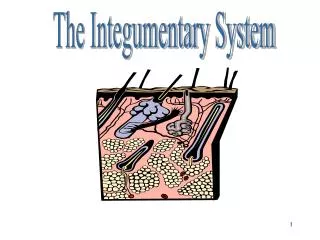
Size of the Integument • The integument is the largest system of the body: • 16% of body weight • 1.5 to 2 m2 in area
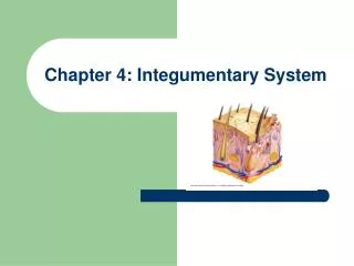
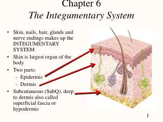
Parts of the Integument • The integument is made up of 2 parts: • cutaneous membrane (skin) • accessory structures
Parts of the Integumentary System Figure 5–1
Parts of the Cutaneous Membrane • Outer epidermis: • superficial epithelium (epithelial tissues) • Inner dermis: • connective tissues
Accessory Structures • Originate in the dermis • Extend through the epidermis to skin surface: • hair • nails • multicellular exocrine glands
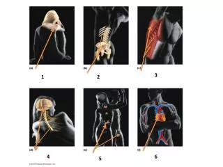
Functions of Skin • Protects underlying tissues and organs • Excretes salts, water, and organic wastes (glands) • Maintains body temperature (insulation and evaporation)
Functions of Skin • Synthesizes vitamin D3 • Stores lipids • Detects touch, pressure, pain, and temperature
What are the main structures and functions of the epidermis?
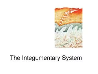
Epidermis • Avascular stratified squamous epithelium • Nutrients and oxygen diffuse from capillaries in the dermis
Organization of the Epidermis Figure 5–2
Layers of the Epidermis • From basal lamina to free surface: • stratum germinativum • stratum spinosum • stratum granulosum • stratum lucidum • stratum corneum
Stratum Germinativum • The “germinative layer”: • has many germinative (stem) cells or basal cells • is attached to basal lamina by hemidesmosomes • forms a strong bond between epidermis and dermis
Structures of Stratum Germinativum • Epidermal ridges (e.g., fingerprints) • Dermal papillae (tiny mounds): • increase the area of basal lamina • strengthen attachment between epidermis and dermis
Ridges and Ducts Figure 5–4
Cells of Stratum Germinativum • Merkel cells: • found in hairless skin • respond to touch (trigger nervous system) • Melanocytes: • contain the pigment melanin • scattered throughout stratum germinativum
Stratum Spinosum • The “spiny layer”: • produced by division of stratum germinosum • 8–10 layers of keratinocytes bound by desmosomes • cells shrink until cytoskeletons stick out (spiny)
Cells of Stratum Spinosum • Continue to divide, increasing thickness of epithelium • Contain Langerhans cells, active in immune response
Stratum Granulosum • The “grainy layer” • Stops dividing, starts producing: • keratin: • a tough, fibrous protein • makes up hair and nails • keratohyalin • dense granules • cross-link keratin fibers
Cells of Stratum Granulosum • Produce protein fibers • Dehydrate and die • Create tightly interlocked layer of keratin surrounded by keratohyalin
Stratum Lucidum • The “clear layer”: • found only in thick skin • covers stratum granulosum
Cells of Stratum Lucida • Flat • Dense • Filled with keratin
Stratum Corneum • The “horn layer”: • exposed surface of skin • 15 to 30 layers of keratinized cells • water resistant • shed and replaced every 2 weeks
Keratinization • The formation of a layer of dead, protective cells filled with keratin • Occurs on all exposed skin surfaces except eyes
Skin Life Cycle • It takes 15–30 days for a cell to move from stratum germinosum to stratum corneum
Skin Color • Skin color depends on: • the pigments carotene and melanin • blood circulation (red cells)
Carotene • Orange-yellow pigment • Found in orange vegetables • Accumulates in epidermal cells and fatty tissues of the dermis • Can be converted to vitamin A
Melanin • Yellow-brown or black pigment • Produced by melanocytes in stratum germinativum • Stored in transport vesicles (melanosomes) • Transferred to keratinocytes
Function of Melanocytes • Melanin protects skin from sun damage • Ultraviolet (UV) radiation: • causes DNA mutations and burns which lead to cancer and wrinkles Skin color depends on melanin production and not the number of melanocytes
Melanocytes Figure 5–5
Capillaries and Skin Color • Oxygenated red blood contributes to skin color: • blood vessels dilate from heat, skin reddens • blood flow decreases, skin pales
Cyanosis • Bluish skin tint • Caused by severe reduction in blood flow or oxygenation
Illness and Skin Color • Jaundice: • buildup of bile produced by liver • yellow color • Addison’s disease: • and other diseases of pituitary gland • skin darkening
Illness and Skin Color • Vitiglio: • loss of melanocytes • loss of color
Vitamin D • Epidermal cells produce cholecalciferol(vitamin D3): • in the presence of UV radiation • Liver and kidneys convert vitamin D into calcitriol: • to aid absorption of calcium and phosphorus
Epidermal Growth Factor (EGF) • Is a powerful peptide growth factor • Is produced by glands (salivary and duodenum) • Is used in laboratories to grow skin grafts
Functions of EGF • Promotes division of germinative cells • Accelerates keratin production • Stimulates epidermal repair • Stimulates glandular secretion
The Dermis • Is located between epidermis and subcutaneous layer • Anchors epidermal accessory structures (hair follicles, sweat glands) • Has 2 components: • outer papillary layer • deep reticular layer
The Papillary Layer • Consists of areolar tissue • Contains smaller capillaries, lymphatics, and sensory neurons • Has dermal papillae projecting between epidermal ridges
The Reticular Layer • Consists of dense irregular connective tissue • Contains larger blood vessels, lymph vessels, and nerve fibers • Contains collagen and elastic fibers • Contains connective tissue proper
Characteristics of Dermis • Strong, due to collagen fibers • Elastic, due to elastic fibers • Flexible (skin turgor)
Dermal Circulation Figure 5–8
Arteries • Cutaneous plexus: • a network of arteries along the reticular layer • Papillary plexus: • capillary network from small arteries in papillary layer
Veins • Venous plexus: • capillary return deep to the papillary plexus • Contusion: • damage to blood vessels resulting in “black and blue” bruising
Nerves • Nerve fibers in skin control: • blood flow • gland secretions • sensory receptors • Tactile disks monitor Merkel cells
What are the structures and functions of the subcutaneous layer?
The Hypodermis • The subcutaneous layer or hypodermis: • lies below the integument • stabilizes the skin • allows separate movement
Structure of the Hypodermis • The subcutaneous layer is: • made of elastic areolar and adipose tissues • connected to the reticular layer of integument by connective tissue fibers