Download
1 / 41
470 likes | 2.39k Views
Physical Examination 2 nd Affiliated Hospital China Medical University 内科 郑长青. What is physical examination? Physical examination is a fundamental examining method, it is proceeded by the sense organs such as eyes, ears, nose and hands or simple tools –stethoscope and plexor. .

E N D
Physical Examination2nd Affiliated Hospital China Medical University内科 郑长青
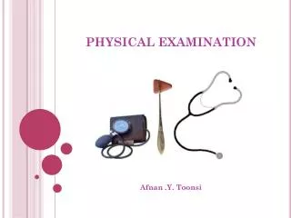
What is physical examination?Physical examination is a fundamental examining method, it is proceeded by the sense organs such as eyes, ears, nose and hands or simple tools –stethoscope and plexor.
The basic methods of physical examination 1 inspection 2 palpation 3 percussion 4 auscultation 5 smelling
1 Inspectionincludes a general view of the patient’s mental status development nutrition posture body movement gait facial expression complexion
For example pallor anemia exophthalmus hyperthyroidism cyanosis(lips) mitral face moon face cushing syndrome spider angioma liver cirrhosis barrel chest pulmonary emphysema
Gastric type pylorus obstruction Abdomenal respiratory movement normal men children disappear acute peritonitis lower extremity edema right heart failure skeleton and joints deformity
2 Palpationmainly used in abdominal examination mass: location size contour consistency mobility tenderness pulsation
The methods of palpationLight palpation Deep Palpation deep slipping palpation bimanual palpation deep press palpation ballottement
The methods of palpation light palpation abdominal muscle tensity abdominal tenderness
Deep Palpationdeep slipping palpation ---deep mass bimanual palpation ---liver spleen kidney deep press palpation ---tenderness point ballottement
bimanual palpation liver and spleen
Cholecystic pointMurphy’s sign ---acute cholecystitis Appendix pointMcBurney point ---acute appendicitis
rebound tenderness ---acute peritonitis ballottement ---liver enlargement ---splenomegaly with massive ascites
Notice:patient in supine position ask patient flex his thighs and knees, tell patient relax his abdomenal muscles. Doctor stands at the right side of patient, warm hand, use your palmar aspect of finger, examining gently and lightly, from superficial to deep.
3 Percussionlungs ---marginheart ---size and shape liver ---upper margin ascites ---shifting dullness
Percussion methodsThere are two methods that may be used for percussion Indirect percussion Direct percussion
Indirect Percussion pleximeter ---usually the middle finger of left hand plexor ---usually the middle finger tip of right hand
Direct percussionThis method can be done by striking chest with the palmar aspect of right hand or the tips of all of the fingers held firmly together, mainly used to examine massive fluid in thoracic cavity.
Percussion SoundsResonance DullnessTympany Flatness Hyperresonance
ResonanceThe sound heard normally over lungs moderately low in pitch
Dullness:This is a short high pitched and is not loud. The sounds heard over heart or liver which are covered with lung tissues or during pneumonia.
Tympany:Somewhat similar to the sound of a drum. The sound in loud intensity, it results from air in a chamber such as stomach and bowel or in pneumothorax
Flatness:Flatness will be present when there is an extensive pleural effusion or over a solid organ such as the liver and heart
HyperresonanceIt is lower pitch than normal resonance, it is heard normally in children and pulmonary emphysema in adult
4 Auscultationthere are two methods of auscultation: direct auscultation with the ear; indirect auscultation with the stethoscope.
by these two methods we can listen to the sounds produced from heart lungs and abdomen or the blood vessel note
There are two principal type of stethoscope the bell and the diaphragm the diaphragm type of chestpiece is more suitable to listen to the high pitched tones
such as the murmur of aortic regurgitation the bell type is more suitable to listen to the low pitched rumble such as the murmur of mitral stenosis
Notice:The stethoscope should be placed firmly against the chest wall or other part of the body to exclude extraneous sounds
5 Smellingto identify the unusual odor which produced from patient such as the odor of sweat sputum pus fluid vomitus stool urine breath