Evo-Devo
Evo-Devo. Developmental Patterns and Evolutionary Patterns Studying Cell Patterns. 10.4 Developmental Patterns and evolution. Similar developments indicate evolutionary relationships “Ontogeny recapitulates Phylogeny” Ernst Haeckel Dev’t is a replay of ancestry.
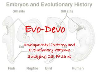

Evo-Devo
E N D
Presentation Transcript
Evo-Devo Developmental Patterns and Evolutionary Patterns Studying Cell Patterns
10.4 Developmental Patterns and evolution Similar developments indicate evolutionary relationships “Ontogeny recapitulates Phylogeny” Ernst Haeckel Dev’t is a replay of ancestry
How is morphogenesis determined? What are the literal instructions?
10.6 Birth Defects • Causes: • defective/mutant gene • environmental factors acting on developmental genes • Polydactyl- extra digits • Spina bifida- part of neural tube does not close completely • Anencephaly- brain does not completely develop- exposed brain degenerates and top of skull fails to form
How do we know? • DNA RNA proteins (responsible for phenotype/what we see expressed as a trait) • All DNA is present in ALL cells • Not all DNA is being “read” • there4, only certain proteins are made • RNA is the intermediary between DNA and protein • If RNA is present, that is the proof that the DNA is being “read”
DNA-RNA Hybridization • Shows what DNA/genes are being expressed • DNA is made single-stranded and tagged with flourescence • Labeled DNA is mixed with embryonic cells • Under microscope, can see if DNA bound RNA
10.8 genetic equivalence of differentiating cells • What happens to unused genes after cell differentiation? Two theories: 1. Selective- Gene- Loss Hypothesis • differentiating cells lose some genes 2. Gene Equivalence Hypothesis • All cells have same genes but only some are active during differentiation. Genes are not lost- just not expressed.
Which Idea is Correct? • 2 experiments: • 1952- Briggs and King (Fig. 10.19) • Blastocyst v. skin cell • Followed soon after by John Gurdon’s Experiment • Tadpole v skin cell • CONCLUSION: • All cells are genetically equal. • No genes are lost. • Differentiation restricts the expression of some genes.
Determination Many embryonic cells are committed to their fate long before differentiation ever begins. Proteostomes tend to be earlier deuterostomes Snail vs. frog exp’ts Sometimes cells determine independently Other cells communicate and affect each other’s determination http://www.youtube.com/watch?v=LsxBhBsLNg8 10.9 Determination and Differentiation
2 separated frog cells- each develop into tadpole • Snail cells were irreversibly determined already- would not develop into whole snail *In the frog the cells adjust. The snail could not adjust because cell fate determined early in development WHY? Proteins & RNA distribute unevenly- separated into different cells through cleavage
10.11 Cell-Cell Interactions:Embryonic Induction • Differentiation is more flexible in vertebrate embryos • Demonstrated by Spemann and Mangold, 1920 • Cells moved around in embryo responded to other nearby cells and altered determination • Example: skin ectoderm cells moved to dorsal region above notchord became neural tube instead of skin • This process is called Embryonic Induction • Early blastula but not late gastrula
10.11 Cell- Cell Interaction: Role of Notochord • Spemann and Mangold • Demonstrated that the notochord is the source of the inducing signal • Experiment: tissue determined to become notochord was transferred to another organism 2nd notochord grew second larva formed! • Later experiments: • Cell contact not required • Inducing proteins are released: chordin and noggin • Chordin- notochord formation, anterior-posterior axis • Noggin- formation of dorsal structures, ventral ectoderm becomes nerve tissue