Download
1 / 31
310 likes | 357 Views
Learn about the classification and identification of microorganisms, prepare Gram stains, and explore bacterial characteristics and shapes in this comprehensive lab on bacteria.

E N D
Purpose of lab 13: • Review classification within the biological world • Understand characteristics used to identify microorganisms • Learn how to prepare a Gram stain
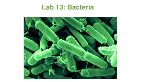
Three Lineages of Life: Domain Bacteria All living organisms are classified into 1 of 3 domains: • Prokaryotes: No true nucleus No membrane-bound organelles • Cell Wall composed of peptidoglycan • Reproduce asexually by budding and fission • Very small (1 - 10 µm)
Three Lineages of Life: Domain Archaea • Prokaryotes: • No true nucleus • No membrane-bound organelles • NO peptidoglycan in cell wall • Reproduce asexually by budding and fission • Very small (1 - 10 µm) • “Extreme” environments (high temperatures)
Identification of Bacteria • Bacteria: • Small in size • Lack discernable internal features • Methods of identifying bacteria: • Macroscopic examination: • Colony color, shape, and odor • Microscopic examination: • Cell shape • Cell surface
Bacterial ID : Microscopic Examination : Cell Shape Unflagellated bacteria are classified according to 1 of 3 major shapes:
Microscopic Examination: Cell Shape: Coccus Single Coccus Diplococcus (paired) Coccus Streptococcus (chain) Staphylococcus
Microscopic Examination: Cell Shape: Bacillus Single Bacillus Single Bacillus (fusiform) Bacillus Streptobacillus (chain)
Microscopic Examination: Cell Shape: Spirillum Single Spirillum Spirillum Single Spirillum
I. EUBACTERIA Examine bacteria cultures both macroscopically and microscopically. characteristics Whole Shape Of Colony • Edge/Margin Of Colony • Color • Opacity Of Colony Transparent (clear), opaque, translucent (almost clear, but distorted vision–like looking through frosted glass), iridescent (changing colors in reflected light) • Elevation Of Colony • Surface Of Colony Smooth, glistening, rough, dull (opposite of glistening), rugose (wrinkled) • Consistency Butyrous (buttery), viscid (sticks to loop, hard to get off), brittle/friable (dry, breaks apart), mucoid • Emulsifiability Of Colony Is it easy or difficult to emulsify? Does it form a uniform suspension, a granular suspension, or does not emulsify at all? • Odor Absent or present? If it has an odor, what does it smell like? • A. Macroscopic Observations of Bacteria Colonies • Pick two colonies from the plates provided, each from a different source. • Prepare a table (Table I) that summarizes these characteristics for each colony
I. EUBACTERIA C. Prepared Microscope Slides of Bacteria Prepared slides of various types of bacteria will be made available. You should examine them, noting and diagramming their characteristics. Bacillus subtilis Staphylococcus epidermidis Escherichia coli Neisseria sicca
Bacterial ID : Microscopic Examination : Cell Surface Nearly all prokaryotes have cell walls • External to their cell membrane • Domain Bacteria (“eubacteria”), PEPTIDOGLYCAN • GRAM STAIN: • One of the most valuable tools for identifying eubacteria • Separates eubacteria into 2 groups based on differences in their cell walls Gram Positive Gram Negative
Gram staining a Important Technique • A staining technique used to classify bacteria • Bacteria are stained with gentian violet and then treated with Gram's solution • After being decolorized with alcohol and treated with safranine and washed in water, those that retain the gentian violet are Gram-positive and those that do not retain it are Gram-negative
Microscopic Examination : Cell Surface : Gram Positive • Gram-positive • Large amounts of peptidoglycan in cell wall • Peptidoglycan traps the violet dye (crystal violet) • These cells stain purple
Microscopic Examination : Cell Surface : Gram Negative Gram-negativeThin peptidoglycan It’s located in a periplasmic gel between the cell membrane and an outer membrane The violet dye is easily rinsed out The red counter-stain (safranin) is retained These cells stain pink
Organizing the Staining Bottles Dr.T.V.Rao MD
1. Place one needle of solid bacterial growth or two loops of liquid bacterial growth in the center of a clean slide.
2. If working from a solid medium, add one drop (and only one drop) of water to your specimen with a water bottle. If using a broth medium, do not add the water.
3. Now, with your inoculating loop, mix the specimen with the water completely and spread the mixture out to cover about half of the total slide area.
4. Place the slide on a slide warmer and wait for it to dry. The smear is now ready for the staining procedure.
Gram-staining Procedure • Gram-staining is a four part procedure which uses certain dyes to make a bacterial cell stand out against its background. The specimen should be mounted and fixed on a slide before you proceed to stain it. • The reagents you will need to successfully perform this operation are: • Crystal Violet (the Primary Stain) • Iodine Solution (the Mordant) • Decolorizer (ethanol is a good choice) • Safranin (the Counterstain) • Water (preferably in a squirt bottle)
Before starting, make sure that all reagents, as well as the squirt-bottle of water, are easily accessible because you won't have time to go get them during the staining procedure. Also, make sure you are doing this near a sink because it can get really messy. Wear a lab coat.
STEP 1: • Place slide on rack. • Flood with crystal violet (~1 min) • Let stand 60 seconds • Rinse with water (5 sec)
STEP 2: Flood with iodine (~1 min) Rinse with water (5 sec) At this point, the specimen should still be blue-violet. Proceed to STEP 3.
STEP 3: Add the ethanol dropwise until the blue-violet color is no longer emitted from your specimen Rinse with water (5 sec)
STEP 4: Rinse with water (5 sec) Flood with safranin(~1 min) Let stand 60 seconds Gram positive cells will remain blue-violet in appearance. Gram negative bacteria take on a pink color ..
After you have completed steps 1 through 4, you should blot the slide gently with bibulous paper or allow it to air dry before viewing it under the microscope. DO NOT RUB THE SMEAR!
Gram staining not a fool proof procedure • Excessive heat during fixation • Low concentration of crystal violet • Excessive washing between steps • Insufficient iodine exposure • Prolonged decolourization • Excessive counterstaining
Part II: CYANOBACTERIA Observe (400X), draw and label: C. Anabaena B. Oscillatoria A. Gleocapsa