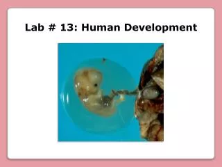
Lab # 13: Human Development
Lab # 13: Human Development. Zona pellucida. Zona pellucida. Female pronucleus. Ovulation of mature ( graafian) follicle. Corona radiata. Corona radiata. Oocyte at Ovulation. It is composed of several layers of granulosa cells. First polar body. Egg membrane . Egg.

Lab # 13: Human Development
E N D
Presentation Transcript
Zona pellucida Zona pellucida Female pronucleus Ovulation of mature (graafian) follicle Corona radiata Corona radiata Oocyte at Ovulation It is composed of several layers of granulosa cells First polar body Egg membrane Egg It is a layer of glyco-protein gel secreted by granulosa cells around the oocyte Secondary oocyte (arrested in metaphase of meiosis II) If not fertilized If fertilized Oocyte dies It completes meiosis II Second polar body (dies) Egg Zygote Embryo
1 2 3 4 Rejected sperm Fertilization membrane It is the exocytosis of the acrosome, releasing the enzymes needed to penetrate the egg Acrosomal reaction • Two acrosomal enzymes are released: Cortical reaction • Hyaluronidase, which digests the hyaluronic acid that binds granulosa cells together • Acrosin, a protease similar to trypsin • When a path has been cleared, a sperm binds to the zona pellucida Cortical granules
The Preembryogenic Stage 1- Cleavage 2- Implantation It comprises the first 16 days of development, culminating with the existence of an embryo. 3- Embryogenesis It refers to the mitotic divisions that occurs in the first 3 days, while the conceptus migrates down the uterine tubes Cleavage: Morula (72 hours) It is a solid ball of 16 cells that resemble a mulberry Blastomeres Zygote • The morula lies free in uterine cavity for 4-5 days and divides into a 100 cells or so 2-celled stage (30 hours) 4-celled stage 8-celled stage Egg pronucleus Blastocyst Sperm pronucleus • The zona pellucida dis-integrates and releases conceptus: blastocyst Fertilization (0 hours) Migration of the Conceptus Implanted blastocyst (6 days)
2- Implantation (about day 6) The trophoblast secrets human chorionic gonadotropin (HCG), which stimulates the corpus luteum to secret estrogen and progesterone (it suppresses menstruation) Blastocyst: Blastocoel Trophoblast Inner cell mass or Embryoblast Endometrium: Epithelium Endometrial gland (6-7 days) The blastocyst attaches to uterine wall 6 days after ovulation, usually on the fundus or the posterior wall of the uterus Implantation: It is the process of attachment to uterine wall that begins when blastocyst adheres to the endometrium
3- Embryogenesis (by the end of 2nd week) Amniotic cavity Embryogenesis: Ectoderm It is the arrangement of the embryoblast into three primary germ layers: ectoderm, mesoderm, and endoderm Mesoderm Endoderm The embryoblast separates slightly from the trophoblast and creates a narrow space between them: the amniotic cavity Yolk sac Once the three primary germ layers are formed, embryogenesis is complete and the individual is considered an embryo. It is about 2 mm long and 16 days old.
The ectoderm and endoderm are epithelia composed of tightly joined cells. The mesoderm is a more loosely organized tissue which differentiates into a loose fetal connective tissue called mesenchyme
Amniotic cavity Formation of Embryonic Membranes and Placenta Yolk sac Chorion Chorionic villi Amnion Placental sinus • They are extensions of syncytiotrophoblast into endometrium by digestion and growth of “roots” of tissue 16 days • They are lacunae filled with maternal blood that merge and surround villi
Placenta and Umbilical Cord As the placenta grows: • The villi grow and branch and their surface area increases • The membrane becomes thinner and more permeable • The placental conductivity (the rate at which substances diffuse through the membrane) increases • Materials diffuse from the side of the membrane where they are more concentrated to the side where they are less concentrated • Oxygen and nutrients pass to the fetal blood. Fetal wastes pass the other way and are eliminated by the mother • The placenta is also permeable to nicotine, alcohol, and most other drugs that may be present in the maternal blood stream