Download
1 / 80
820 likes | 1.02k Views
Chapter 23— The Urinary System. Ch. 23 Study Guide. Critically read 23.1 to right before the 23.7 (Urine Storage and Elimination) section (pp. 905-931) Comprehend Terminology (those in bold) Study-- Figure questions, Think About It questions, and Before You Go On (section-ending) questions

E N D
Ch. 23 Study Guide Critically read 23.1 to right before the 23.7 (Urine Storage and Elimination) section (pp. 905-931) Comprehend Terminology (those in bold) Study-- Figure questions, Think About It questions, and Before You Go On (section-ending) questions Do end-of-chapter questions: Testing Your Recall— 2-10, 12-17, 19-20 True or False– 1-9 Testing Your Comprehension-- #1-2 2
§ Major Nitrogenous Wastes Ammonia: very toxic; from amino group in the a.a. Urea: less toxic; converted from ammonia in the __________ Uric acid: nucleic acid catabolism Creatinine creatine phosphate catabolism
§ Major Nitrogenous Wastes BUN (Blood Urea Nitrogen) • Measure the amount of ________ in your blood • Why is it done? • Disorders: • Azotemia: an abnormally elevated BUN is called; may indicate renal insufficiency • Uremia: toxic effects as wastes (urea) accumulate in the blood; patients with renal failure • Symptoms– vomiting, diarrhea, cardiac arrhythmia etc. • Treatment--Hemodialysis
§ Major kidney functions (1) • Eliminate wastes & foreign compounds • Major nitrogenous wastes? • Major foreign cpds? • Maintain blood volume and electrolyte (ion) concentration • For example, water balance during exercise • Another example, [K+] and ectopic focus
§ Major kidney functions (2) • Produce hormones— two major ones? • Detoxifies free radicals and drugs • In times of starvation-- gluconeogenesis (makes glucose from amino acids or fats)
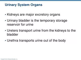
§ The urinary system • Kidneys (a pair) • Functions-- • The ureter(a pair) • The single urinary bladder • A muscular sac for _________________ • The urethra • Draining urine to the outside • In females vs. in males (length and external orifice) + proper toilet habits Figure 23.1
§ Gross Anatomy of Kidney • Renal cortex: outer 1 cm; extensions of the cortex called renal columns • Renal medulla: inner zone; renal columns divide the medulla into 6-10 renal pyramids (each blunt point called renal papilla) • The renal papilla is nestled in a cup called a minor calyx major calyx • Lobe of kidney: pyramid and it’s overlying cortex Fig. 23.4 + a practice figure
Figure 23.5a § Renal circulation-- C A B
Path of Blood Through Kidney • Renal artery: into segmental artery and then A--interlobararteries(up renal columns, between lobes) B--arcuatearteries (over pyramids) C--interlobulararteries (up into cortex) afferentarterioles glomerulus (cluster of capillaries) efferentarterioles (near medulla vasa recta) peritubular capillaries interlobular veins arcuate veins interlobar veins • Renal vein
A. B.
Two kinds of Nephrons, depending upon locations • Cortical nephrons (85%) • short nephron loops of Henle • efferent arterioles branch off into peritubular capillaries • Juxtamedullary nephrons (15%) • Where? • very long nephron loops-- • Vasa Recta– Efferent arterioles descend into the medulla and give rise to Vasa Recta instead of peritubular capillaries. • The capillaries of the vasa recta lead into venules that empty into the interlobular and arcuate veins
§ The Nephron (2) • How many nephrons in each kidney? Answer: ___________ • The nephron are blood-processing units and each one of them is a functional unit of the kidneys • Vascular and tubular parts of the nephron • Vascular parts first--Figure 23.6, 23.7
4. Efferent arteriole 3. Glomerulus 2. Afferent arteriole 1. Interlobular Artery 6. Interlobular vein 5. Peritubular capillaries To renal pelvis
§ Vascular part of the Nephron (3) • The renal artery (. . . interlobular artery)– • Afferent arteriole– • supplies each nephron and delivers blood to the glomerulus • The glomerulus– cluster of capillaries (1st set of capillary in each nephron); function?
§ Vascular part of the Nephron (4) • The efferent arteriole— • Where the glomerular capillaries rejoin • The peritubular (2nd set of) capillaries-- • Impt in exchanges between blood and ______________ • The renal veins-- • The major blood vessels leave the kidney --------------------------------------------------------------- • Tubular parts of the nephron– @Fig. 23.8
1. Bowman’s capsule 4. Distal tubule 2. Proximal tubule Glomerulus 5. Collecting duct Artery Vein Cortex Medulla 3. Loop of Henle (nephron loop) To renal pelvis
§ Tubular part of the Nephron (5) • A hollow tube formed by a single layer of epithelial cells; They are, in order: • Bowman’s (Glomerular) capsule– • Cup-shaped; double-wall invagination • Surround each __________ • Proximal tubule– closest to Bowman’s capsule • Lies entirely within the cortex
§ Tubular part of the Nephron (6) • The loop of Henle (nephron loop)– • Forms a U-shaped loop • The distal tubule– • most distant from the capsule; lies entirely within the ____________ • Collecting tubule/duct— • drains fluid from up to 8 nephrons Figure 23.8
Figure 23.8b 1. 4. 2. 5. 3. ID parts (1-5) of the nephron.
§ Three urine forming processes 1.-- Glomerular filtration • From the glomerulus into Bowman’s (glomerular) capsule 2A.--Tubular reabsorption • From the tubular lumen into ___________ 2B.--Tubular secretion • From the peritubular capillaries into the __________________ 3.-- Water conservation Figure 23.9
Different names (fluid in renal tubules) in different areas: 1. Glomerular filtrate (in the capsular space) 2. Tubular fluid (proximal tubule to distal tubule) 3. Urine (collecting duct and beyond)
§ 1. Glomerular filtration • Def.– filtering blood by forcing small molecules into the Bowman’s capsule • What in the filtrate? • Small molecules can pass— • Large molecules cannot— • Mechanism? ATP? • What is the major force? Glomerular blood hydrostatic pressure (BHP)
§ 1. Glomerular filtration (cont.) • Layers of the glomerular filtration mem. • 1-Fenestrated endothelium of capillaries • Large pores (100x more permeable) • Molecules can pass– • 2-(Acellular) basement mem. • Collagen & glycoproteins • Function-- • 3a-Filtration slits; present in inner layer of the Bowman’s capsule-- podocytes (3b) bear many foot processes (pedicles) Figure 23.10 (a-d)
Afferent arteriole Efferent arteriole Glomerulus Bowman’s capsule Lumen of glomerular capillary Endothelial cell Lumen of Bowman’s capsule Basement membrane (see next slide) Outer layer of Bowman’s capsule Podocyte foot process Inner layer of Bowman’s capsule (podocytes) Proximal convoluted tubule
3b. Podocyte & foot process 3a.Filtration slits 2. Basement membrane 1. Capillary pore
1b. Endothelial cell 1a. Capillary pore Lumen of glomerular capillary 2. Basement membrane Lumen of Bowman’s capsule (capsular space) 3a.Filtration slit 3b. Podocyte foot process
A. Endothelial cell Lumen of glomerular capillary B. Lumen of Bowman’s capsule Filtration slit C.
§ 1. Glomerular filtration (cont.) Disorders: • Albuminuria– also called proteinuria; presence of ________ in the urine • Criteria: >250 mg/day: pathological • Hematuria– presence of ______ in the urine
§ 2. Tubular reabsorption • Def. reclamation process to move molecules back into the blood • Goal: to move molecules from tubular lumen to the peritubular capillaries (or vasa recta) Table x & Figure y
§ 2.Tubular reabsorption (cont.) • What are reabsorbed? • All the glucose, vitamins, and . . . • How efficient? • Glucose— no glucose escapes • Water— 180 L filtrate to 1-2 L of urine/day • Analogy— Clean out a cluttered drawer
III. Urine Formation; 2A. TUBULAR REABSORPTION– in the proximal convoluted T.
§ 2. Tubular reabsorption (cont.) Two examples—Na+, water in proximal convoluted tubules and beyond • --1st example– sodium reabsorption • Where are sodium ions been reabsorbed? Most of the tubule Exception is the descending limb of the loop of Henle • Routes taken: both transcellular and paracellular routes
§ 2. Tubular reabsorption (cont.) • Mechanisms of sodium reabsorption— • A--symport proteins (channels)— • B--Na+-H+ antiport— • C--Na+-K+ pumps– basal and lateral membrane • D– Paracellular route-- Figures 23.16
§ 2 Tubular reabsorption (cont.) • --2nd example– water reabsorption Locations? All the renal tubule; however, 2/3 occurs in PCT • Mechanisms— Via water channels (aquaporins) Between cells Water moves into blood plasma Figures 23.16 23-48
§ Reabsorption Limit • Def.-- A limit to the amount of solute the renal tubule can reabsorb • Why? Limited no. of transport proteins • Tm= Transport maximum; example-- • Glucose’s Tm is 320 mg/min • Glucose normally enters the renal tubule at 125 mg/min; will all of it be reabsorbed? • Threshold of glucose in the plasma– 220 mg/dL (= 220mg/100mL); begin to see glucose in the urine called glycosuria • Untreated diabetes mellitus patients– 400 mg/dL (plasma glucose) 23-50