The Cardiac Cycle Chapter 3
210 likes | 423 Views
The Cardiac Cycle Chapter 3. Ara G. Tilkian, MD, FACC Instructor Patricia L. Thomas, MBA, RCIS. The Cardiac Cycle. Atrial and Ventricular Diastole and Systole The sounds of the Heart The valves and papillary Muscles Valvular Pathophysiology. Atrial & Ventricular Diastole.

The Cardiac Cycle Chapter 3
E N D
Presentation Transcript
The Cardiac Cycle Chapter 3 Ara G. Tilkian, MD, FACC Instructor Patricia L. Thomas, MBA, RCIS
The Cardiac Cycle • Atrial and Ventricular Diastole and Systole • The sounds of the Heart • The valves and papillary Muscles • Valvular Pathophysiology
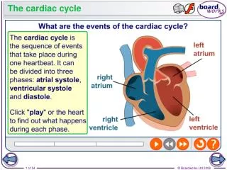
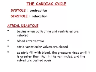
Atrial & Ventricular Diastole • Phase of Cardiac Cycle (see 3.1 A) • Atrial Diastole • Venous blood enters RA via SVC & IVC • Pulmonary Blood enters the LA via the 4 PV
Atrial & Ventricular Diastole • Ventricle Diastole-two phases • #1 Early passive filling (both atria/ventricles are relaxed • #2 Late active filling (atrial systole or A-Kick)(see 3.1 B) • 3rd and 4th heart sounds (S3 & S4) • 3rd HS the initial rapid filling phase early in diastole • 4th HS the final filling phase at the end of diastole
Atrial and Ventricular Systole • Atrial Systole • Occurs in response to depolarization • Contraction occurs toward the end of ventricular diastole (atrial-kick) before the ventricles contract • Electrical impulse traveling to the ventricles is normally delayed momentarily in the AV node premitting the atrial contraction to augment ventricular filling.
Atrial and Ventricular Systole • Ventricular Systole • Occurs in response to the depolarization wave within the ventricles the QRS wave • Venous blood is propelled from the RV the the PA for oxygenation • Oxygenated blood is propelled from the LV through the AO to systemic circulation • Onset of ventricular systole the AV and PV are still closed. (isovolumic ventricular contraction) (see 3-1 C) • When the aortic and pulmonic valves open (rapid ejection)
Isovolumic Ventricular Contraction, Beginning of Ventricular Systole
The Sound of the Heart • S1 closure of the Mitral & Tricuspid valves • S2 closure of the Aortic & Pulmonic • S3 passive ventricular filling sound (early diastole) • S4 active ventricular filling sound (late diastole)
The First Heart /Ejection Sounds • Mitral valve closure closely followed by tricuspid valve closure • Closure occurs when the onset of ventricular systole abruptly raises the ventricular pressure above that of the atria • The aortic valve opens (AO) followed by the opening of the pulmonic valve (PO) • The opening of the two semilunar valves is normally not heart • A diseased aortic/pulmonic valve may produce an ejection sound
The Second Heart Sound • Aortic valve closure (AC) closely followed by pulmonic valve closure (PC) • The two valves close when the systolic ejection into the aorta/pulmonary artery declines and rising pressure in these great vessels exceeds the pressures in the respective ventricles, reversing the flow and causing the closure of their valves. • Isovolumic relaxation is after the closure of the AV/PV but before opening of the MV/TV results in a rapid fall in ventricular pressure • Ventricular = Atrial pressure MV/TV opens silently • If valves are abnormal/stenosed an opening snap (OS) or click may be heard
The Third Heart Sound • Rapid filling of the left ventricle following the opening of the mitral valve • Physiological in young people • Pathological in people with congestive heart failure or ventricular dilatation
The Fourth Heart Sound • Blood being forced into a stiff or noncompliant LV by the atrial contraction • Late diastolic event that should be silent • Atrial diastolic gallop when heard • Stiff ventricles may be caused by hypertension, ischemia, outlet obstruction, or hypertrophic or infiltrative myopathies
The Atrioventricular Valves • Competence of the AV valves depends on the chordae tendineae (CT) • CT are attached to the free edges and ventricular surfaces of the valve cusps • CT are attached to fingerlike projections of muscle tissue from the endocardium called the papillary muscles
The Semilunar Valves • Aortic and Pulmonic valves are called semilunar because of their half-moon shape, a structure that prevents the backflow of blood form the aorta and pulmonary artery into the ventricles.
Valvular Pathophysiology • Mitral/Tricuspid stenosis-when these values are unable to open completely or have a congenitally small opening there is an obstruction to the free flow of blood • Competent- a properly functioning valve • Regurgitant/Insufficient-leaky or stenosed valve
THE ENDOFCHAPTER 3 Tilkian, Ara MD Understanding Heart Sounds and Murmurs, Fourth Edition, W.B. Sunders Company. 2002, pp. 26-33.