Download

1 / 13
130 likes | 257 Views
Fabrication of Percutaneous Transvenous Mitral Annuloplasty Testing Device. Team 19 Kaitlyn Clarke Brittany DePoi Andrew Reynolds Adarsha Selvachandran Client: Dr. Wei Sun. Project Overview.

E N D
Fabrication of PercutaneousTransvenous Mitral Annuloplasty Testing Device Team 19 Kaitlyn Clarke Brittany DePoi Andrew Reynolds AdarshaSelvachandran Client: Dr. Wei Sun
Project Overview • Dr. Sun has requested a testing device that will allow his research team to obtain physical evidence regarding the response of cardiac tissue to implantation of the Percutaneous Mitral Annuloplasty System for the Treatment of Mitral Regurgitation ( aka. “stent bridge constriction device”) • Required project components: • Biological environment regulator • Marker Tracking System • Force Determination System • Ultrasonic Detection Distal Anchor Nitinol Bridge Proximal Anchor
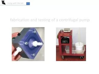
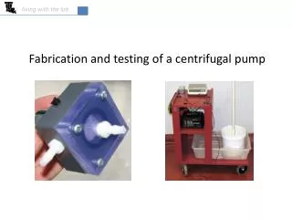
Biological Environment Regulator • Purpose: support heart and simulate naturally occurring environmental conditions such as temperature, pressure, and blood flow • 3 main components: • Water bath • Camera mount and frame • Heart mount
Biological Environment Regulator Water Bath and Camera Mount • Water Bath • Clear cast acrylic sheets • Two separate baths (inner and outer) • Camera Mount • Aluminum • Adjustable
Biological Environment Regulator Heart Mount Aorta Tube • Secure enough to hold heart stable, loose enough to allow heart to move naturally in response to the stent bridge constriction device • Aluminum base • Small hooks to securely hold heart in place • Rigid tube inserted into the ascending aorta, down into the left ventricle • Structural support • Pathway for saline that will control pressure in the mitral valve Base
Marker Tracking System • Purpose: Understand mechanical changes experienced by heart during stent deployment • 15-20 small graphite markers on mitral annulus plane • 2 cameras 30-45 degrees apart to capture image • Calibration Cube in field of view of each camera
Marker Tracking SystemAlgorithm • Direct linear transformation (DLT) MATLAB code • Recreate image with reference to the three-dimensional plane • Determine the 3-D position of each marker • Keep track of each marker and it’s movements from frame to frame • Recognize if a marker leaves the frame of view of one of both cameras • Calculate stress, strain, and deformation
Force Determination System • Purpose: Determine relationship between force applied on the mitral annulus by stent and the decrease in mitral valve prolapse • FlexiForce Sensors • Force measured at specific point as opposed to continuous • Use 6 sensors in an attempt to get more thorough data
Ultrasonic Detection • Purpose: Capture Image of the coronary sinus before and after stent deployment • Ultrasound Machine has 2 probes one probe port • Probe attached to machine will be changed between deployment of stents Distal Stent Proximal Stent
Coronary Sinus Aortic Root Mitral Valve (open)
Mitral Valve (closed) Distance between coronary sinus and valve Coronary Sinus
What’s Next? (We’ve ordered and received Plexiglas/FlexiForce) • Begin to build water bath • Obtain computer from Kewei and begin downloading Vision Software • Build FlexiForce circuit and experiment with sensors to determine the best way to adhere/use them • Test ultrasound to become comfortable with use and to determine the best settings to use