Download
1 / 15
190 likes | 567 Views
Persistent Right 4 th Aortic Arch. James Montgomery, DVM December 8, 2008. Acc # 98847. Barley Referred for vascular ring anomaly 3 months old Male Mixed breed. Acc # 98847. CT (acc # 98848) confirmed right-sided aortic arch Also has left retroesophageal subclavian artery.

E N D
Persistent Right 4th Aortic Arch James Montgomery, DVM December 8, 2008
Acc # 98847 • Barley • Referred for vascular ring anomaly • 3 months old • Male • Mixed breed
Acc # 98847 • CT (acc # 98848) confirmed right-sided aortic arch • Also has left retroesophageal subclavian artery
Persistent Right 4th Aortic Arch (PRAA) • The best documented vascular ring anomaly in dogs and cats (approx. 95% of ring anomalies are PRAA) • Considered to have a familial tendency • Other, less common vascular anomalies include: • Persistent right or left subclavian arteries • Double aortic arch • Persistent right dorsal aorta • Left aortic arch and right ligamentum arteriosum • Aberrant intercostal arteries
PRAA Signalment • Puppies and kittens at time of weaning • German shepherds, Labrador retrievers, and Irish setters appear predisposed • Male and female fairly equally represented
PRAA Clinical Signs • Regurgitation of solid foods at weaning • Weight loss with failure to thrive despite a good appetite • Moist cough, dyspnea, fever – aspiration pneumonia common
Diagnostics • Survey thoracic radiographs • Esophageal body dilation cranial to the heart base • Barium esophagram • Confirm location of esophageal obstruction and severity of esophageal distension • Angiography • Confirm type and location of vascular anomaly prior to surgery • Esophagoscopy • Differentiate intraluminal stricture from extraluminal compression
Radiographic Findings • VD or DV radiographs • Trachea curving to the left rather than the right near the cranial border of the heart • Lateral radiographs • Ventral curvature of the trachea • Marked ventral curvature should prompt a thorough search for additional abnormalities • Retroesophageal left subclavian artery, double aortic arch • Focal narrowing of the trachea cranial to the heart
Differentials • Vascular ring anomalies should be differentiated from: • Congenital idiopathic megaesophagus • Esophageal foreign body • Cricopharyngeal dysphagia
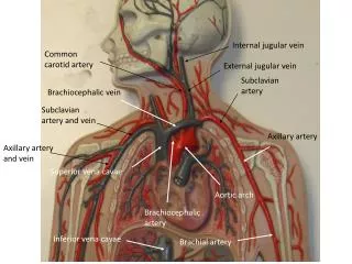
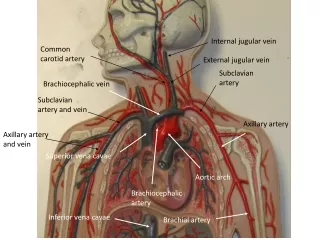
PRAA • Congenital defect in development of the aortic arches • Right 4th aortic arch develops into the aorta rather than the Left 4th aortic arch • Results in esophagus passing to the left of the aorta instead of the right • Esophagus ringed by: • Aorta • Ligamentum arteriosum • Pulmonary trunk • Base of heart
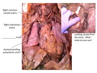
PRAA Buchanan, JVIM 2004
Embryology • Two 3rd aortic arches give rise to the common carotid arteries • Left 4th aortic arch becomes the aorta • 6th aortic arch gives rise to the pulmonary trunk and pulmonary arteries
Associated Vascular Anomalies • Buchanan, JVIM 2004 • 52 dogs with PRAA • 17 (33%) also had retroesophageal left subclavian artery • 6 had PDA • 6 had double aortic arches with atretic left arch • 6 had persistent left cranial vena cava • 3 had a left hemiazygos vein • 2 had PRAA and generalized megaesophagus
Treatment • Surgical ligation and transection of the ligamentum arteriosum • If atretic left aortic arch is present, must transect this as well to relieve esophageal obstruction • Treatment of aspiration pneumonia (if present) • If not diagnosed early, progressive esophageal dilation causes irreversible myenteric nerve degeneration and esophageal hypomotility.
References • Buchanan JW. Tracheal Signs and Associated Vascular Anomalies in Dogs with Persistent Right Aortic Arch. J Vet Intern Med 2004; 18:510-4. • Jergens AE. Diseases of the Esophagus. In Ettinger SJ, Feldman EC, eds. Textbook of Veterinary Internal Medicine, 6thed (St. Louis, MO: Elsevier Saunders, 2005) pp. 1306-7. • Pasquini C, et al. Anatomy of Domestic Animals, 9thed (Pilot Point, TX: Sudz Publishing, 1997) pp. 390-2.