Download
1 / 67
670 likes | 790 Views
This lecture provides an in-depth overview of DNA replication mechanisms in both bacterial (E. coli) and eukaryotic systems (including yeast and human cells). It covers the structure and function of the E. coli replisome, detailing its components and sub-assemblies, as well as the coordination of leading and lagging strand synthesis. The session also explores eukaryotic replisome components, polymerase switching, Okazaki fragment maturation, initiation mechanisms, and termination mechanisms, including fidelity and proofreading processes.

E N D
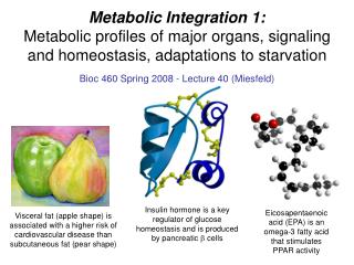
MMG /BIOC 352 The Replisome: DNA Replication in E. coli and Eukaryotes Spring 2006 Scott W. Morrical
Contact Information Scott W. Morrical Given B407 656-8260 Scott.Morrical@uvm.edu
Lecture Outline: Overview of DNA Replication Bacterial systems (E. coli) Eukaryotic systems (yeast/human) The E. coli Replisome Components & sub-assemblies Replisome structure/function Coordination of leading/lagging strand synthesis The Eukaryotic Replisome Polymerase switching Okazaki Maturation Initiation Mechanisms E. coli oriC paradigm Eukaryotic model Termination Mechanisms Tus-Ter Fidelity Mechanisms Proofreading Mismatch repair Processivity Mechanisms: Structure/Function of Sliding Clamps E. colib-clamp Eukaryotic PCNA Structure/Function of AAA+ Clamp Loaders E. coli g-complex Eukaryotic RFC Other AAA+ ATPase Machines
Reference list for this topic: Ref 1: Johnson, A., and O’Donnell, M. (2005) Cellular DNA replicases: components and dynamics at the replication fork. Annu. Rev. Biochem. 74, 283-315. Ref 2: Davey, M.J., Jeruzalmi, D., Kuriyan, J., and O’Donnell, M. (2002) Motors and Switches: AAA+ machines within the replisome. Nat. Rev. Mol. Cell Biol. 3, 826-835. Ref 3: Kong, X.P., Onrust, R., O’Donnell. M. and Kuriyan, J. (1992) Three-dimensional structure of the beta subunit of E. coli DNA polymerase III holoenzyme: a sliding clamp. Cell 69, 425-437. Ref 4: Krishna. T.S., Kong, X.P., Gary, S., Burgers, P.M., and Kuriyan, J. (1994) Crystal structure of eukaryotic DNA polymerase processivity factor PCNA. Ref 5:Jeruzalmi, D., O’Donnell, M., and Kuriyan, J. (2001) Crystal structure of the processivity clamp loader gamma complex of E. coli DNA polymerase III. Cell 106, 429-421. Ref. 6:Bowman, G.D., O’Donnell, M., and Kuriyan, J. (2004) Structural analysis of a eukaryotic sliding DNA clamp-clamp loader complex.
References (cont’d): Ref 7: Mendez, A., and Stillman, B. (2003) Perpetuating the double helix: molecular machines at eukaryotic DNA replication origins. Bioessays 25, 1158-1167. Ref 8: Neylon, C., Kralicek, A.V., Hill, T.M., and Dixon, N.E. (2005) Replication termination in Escherichia coli: structure and antihelicase activity of the Tus-Ter complex. Micr. Mol. Biol. Rev. 69, 501-526 Further Reading: Mammalian DNA mismatch repair. Buermeyer et al. (1999) Annu. Rev. Genet. 33, 533-564. Role of DNA mismatch repair defects in the pathogenesis of human cancer. Peltomaki (2003) J. Clinical Oncology 21, 1174-1179.
DNA Chemistry 5’-end 3’-end Basepair A:T or G:C Backbone Phosphate 2’-deoxy- ribose 5’-end 3’-end
DNA Replication Fork Chemical Inheritance-- DNA Replication • processive • 5’ to 3’ • semi-conservative • semi-discontinuous • high-fidelity
E. Coli Chromosome 1 unique origin of bi-directional replication 10 polar termination sites
Replication Progression of E. coli Chromosome oriC theta structure oriC oriC ter sequences
Replication of Eukaryotic Chromosomes Many different origins on each chromosome firing simultaneously or in a programmed sequence.
DNA Replication Fork • Major Protein Components: • DNA polymerase holoenzyme(s) • -- polymerase • -- proofreading exonuclease • -- sliding clamp • -- clamp loader complex • DNA helicase(s) • Primase • ssDNA binding protein • Other accessory factors needed for correct assembly, processive movement, and fidelity.
Major Components of E. coli Replisome: PolIII-- DNA polymerase III holoenzyme (Pol III) DnaG primase DnaB helicase SSB-- ssDNA-binding protein Plus accessory proteins, loading factors
Replisome Mol. Component Wt. [stoichiometry] Gene (kDa) Function Pol III holoenzyme 791.5 Dimeric, ATP-dependent, processive polymerase/clamp loader Pol III star 629.1 Dimeric polymerase/clamp loader Core 166.0 Monomeric polymerase/exonuclease a [2] dnaE 129.9 5’ --> 3’ DNA polymerase e [2] dnaQ 27.5 3’ --> 5’ exonuclease q [2] holE 8.6 Stimulates e exonuclease g/t complex 297.1 ATP-dependent clamp loader g/t [1/2] dnaX 47.5/71.1 ATPase, t organizes Pol III star and binds DnaB d [1] holA 38.7 Binds b clamp d’ [1] holB 36.9 Stator, stimulates g ATPase in ATP site 1 c [1] holC 16.6 Binds SSB y [1] holD 15.2 Connects c to clamp loader b [2 dimers] dnaN 40.6 Homodimeric processivity sliding clamp Primase [1] dnaG 65.6 Generates primers for Pol III holoenzyme DnaB helicase [6] dnaB 52.4 Unwinds duplex DNA 5’ --> 3’ ahead of the replication fork SSB [4] ssb 18.8 Melts 2o structure in ssDNA, binds clamp loader through c E. coli Replisome Stoichiometries
E. coli g Complex-- ATP-dependent clamp loading activity
Structural Organization of Pol III Holoenzyme
Eukaryotic Replisome Components S. cerevisiae (kDa)H. sapiens (kDa)Function and remarks [S. pombe name] RFCa RFC (277.7)a RFC (314.9)a Pentameric clamp loadera RFC1 (94.9) p140 (128.2) Binds ATP; phosphorylated RFC2 (39.7) p37 (39.2) Binds ATP RFC3 (38.2) p36 (40.6) Binds ATP RFC4 (36.1) p40 (39.7) Binds ATP RFC5 (39.9) p38 (38.5) Binds ATP or ADP PCNAa PCNA (28.9) a PCNA (28.7) 87 kDa a Homotrimeric sliding clamp a Pol d a Pol d (220.2) a Pol d (238.7) a Replicative DNA polymerase a Pol3 (124.6) p125 (123.6) DNA polymerase, 3'-5' exo, binds PCNA; subunit A [S.p. Pol3] Pol31 (55.3) p50 (51.3) Structural subunit; subunit B [S.p. Cdc1] Pol32 (40.3) p66 (51.4) Binds PCNA; subunit C [S.p. Cdc27]; binds Pol a large subunit — p12 (12.4) Structural, stimulates processivity; subunit D [S.p. Cdm1] Pol e a Pol e (378.7) a Pol e (350.3) a Replicative DNA polymerase a Pol2 (255.7) p261 (261.5) DNA polymerase, 3'-5' exo [S.p. Pol2/cdc20] Dpb2 (78.3) p59 (59.5) Binds polymerase subunit [S.p. Dpb2] Dpb3 (22.7) p17 (17.0) Binds Dpb4 Dpb4 (22.0) p12 (12.3) Present in ISW2/yCHRAC chromatin remodeling complex [S.p. Dpb4] Pol a a Pol a (355.6) a Pol a (340.6) a DNA polymerase/primase a Pol1 (166.8) p180 (165.9) DNA polymerase Pol12 (78.8) p68 (66.0) Structural subunit Pri2 (62.3) p55 (58.8) Interacts tightly with p48 Pri1 (47.7) p48 (49.9) RNA primase catalytic subunit aInformation concerns a protein complex.
Eukaryotic Replisome Components (cont’d) S. cerevisiae (kDa)H. sapiens (kDa)Function and remarks [S. pombe name] MCM a MCM (605.6) a MCM (535) a Putative 3'-5' replicative helicase a Mcm2 (98.8) Mcm2 (91.5) Phosphorylated by Dbf4-dependent kinase Mcm3 (107.5) Mcm3 (91.0) Ubiquitinated, acetylated Mcm4 (105.0) Mcm4 (96.6) Helicase with MCM6,7; phosphorylated by CDK; aka Cdc54 Mcm5 (86.4) Mcm5 (82.3) Aka Cdc46; Bob1 is a mutant form Mcm6 (113.0) Mcm6 (92.3) Helicase with MCM4,7 Mcm7 (94.9) Mcm7 (81.3) Helicase with MCM4,6; ubiquitinated RPA a RPA (114) a RPA (100.5) a Single-stranded DNA-binding protein a RPA70 (70.3) RPA70 (70.3) Binds DNA, stimulates Pol a RPA30 (29.9) RPA30 (29) Binds RPA70 and 14, phosphorylated RPA14 (13.8) RPA14 (13.5) Binds RPA30 a Information concerns a protein complex.
Model for Eukaryotic Replisome (Based on E. coli and T4 Phage Models)
Polymerase Switching During Eukaryotic Lagging Strand Synthesis & Okazaki Maturation via RNaseH1 and Fen1/RTH1
Okazaki Maturation Involving Helicase Strand Displacement & Flap Endonuclease Activity of Fen1/RTH1 E. coli: RNA primers removed by 5’ --> 3’ exo activity of DNA polymerase I (Pol I). Simultaneous fill-in with DNA (nick translation rxn) leaves nick that is sealed by ligase.
DNA Replication: Initiation, Termination, & Fidelity Mechanisms Scott Morrical
Initiation of E. coli DNA Replication at oriC-- roles of DnaA Initiator Protein and DnaC Helicase Loader DnaA-- initiator protein oriC-- replicator sequence
Shameless Speculation About Helicase Loading Mechanisms at Eukaryotic Origins
Replication Termination: Direction-specific Termination of DNA Replication by E. coli Tus Protein Bound to a Ter Sequence
E. Coli Chromosome 1 unique origin of bi-directional replication 10 polar termination sites
Replication Fork Arrest by Correctly Oriented Tus-Ter Complex
Alternative Models for the Direction- Specificity of Fork Arrest by Tus-Ter
Replication Fidelity Mechanisms: Spont. Error Frequency Pol 10-4 Pol + exo 10-7 Pol + exo + MMC 10-9 to 10-10
Single base mismatches-- misincorporation by DNA polymerase, missed by proofreading exonuclease. Insertion-deletion loops (IDLs)-- caused by polymerase slippage on repetitive template, gives rise to Microsatallite Instability (MSI).
E. coli Methyl-Directed Mismatch Repair System
Heterodimers of Eukaryotic MutS & MutL Homologs *Note: This is yeast nomenclature. Mlh1 paralogs have different names in yeast and humans. Mlh1-Mlh2 Msh2 Msh3 Mlh1-Mlh3 MutLb Msh2 Msh3 MutSb MutLa Rad1-Rad10 Mlh1-Pms1 Mlh1-Pms1 Mlh1-Mlh3 Msh2 Msh3 Msh2 Msh3 Msh2 Msh6 Msh4 Msh5 MutSa 2-4 b 1 b Non-homologous tail removal in recombination intermediates Insertion/deletion loop (IDL) removal Repair of base-base mismatches Promotion of meiotic crossovers
Sliding Processivity Clamps of DNA Polymerases: X-ray Structure of b-subunit of E. coli DNA Polymerase III
BACKGROUND: Biochemical studies established that beta is essential for processive DNA synthesis by Pol III. Beta exists as a stable dimer in solution. Beta dimer has no intrinsic affinity for DNA, yet in the presence of gamma complex + ATP, beta dimer forms and extremely stable complex with circular, but not with linear, primed DNA molecules. Proposal by O’Donnell & coworkers: b2 is topologically linked, not thermodynamically bound to DNA, and forms a sliding clamp that tethers Pol III. 3’ 5’ 5’ 3’ Pol III core b2
Kong, Onrust, O’Donnell, & Kuriyan (1992) “Three-dimensional structure of the beta subunit of E. coli DNA polymerase III holoenzyme: a sliding DNA clamp”. Cell 69, 425-37. 2.5Å resolution Non-crystallographic 2-fold rotational axis of symmetry perpendicular to face of ring and passing through center of hole. • Highly symmetrical; almost hexagonal symmetry. • Protomers interact head-to-tail in 2 interfaces on opposite sides of the ring.
Space-filling Model of Beta Dimer with B-form DNA Modeled In O.D. ≈ 80 Å, I.D. ≈ 35 Å-- easily accommodates B- or A-form (RNA/DNA hybrid) duplex (~25 Å O.D.) without steric repulsions. Thickness ≈ 34 Å, equal to ~1 full helical repeat of B-form dsDNA.
Connectivity of Beta Subunit • Green (N-terminus) • light blue • purple • red • yellow (C-terminus) • Dimer interfaces are between domains colored green/blue and red/yellow. • Domains are numbered 1,2,3 and 1’, 2’, 3’ in the two monomers.
Unexpected Features of the b2 Structure: Internal symmetry. -- each monomer consists of 3 structural domains of identical chain topology and very similar 3-D structure. -- this was surprising because there are no internal regions of a.a. sequence homology. 2. Each domain is roughly 2-fold symmetric in architecture, with an outer layer of 2 b-sheets providing a scaffold that supports 2 a-helices.
Unexpected Features of the b2 Structure (cont’d): Replication of this motif around a circle (2 subunits x 3 domains/subunit) results in a rigid molecule with 12 a-helices lining the inner surface of the ring, and with 6 “seamless” interlocking b-sheets forming the outer surface. 4. Symmetry of domains gives rise to highly symmetrical and roughly hexagonal star shape of the dimer.
60o rotation about dimer axis superimposes domains. -- different a.a. sequences, but 80% structurally analogous at Ca’s. -- hence the approximately hexagonal symmetry.