Download
1 / 105
1.08k likes | 1.52k Views
The Cardiac Exam. Cheryl Hohe , MSN , APRN, ANP-BC. History. Risk factor’s for heart disease : - HTN -Diabetes -Hyperlipidemia -Family History (be sure to ask for hx of sudden deaths) -Age (>50 for males, >55 for females) -Gender (males and post-menopausal females) -Smoking -Obesity

E N D
The Cardiac Exam Cheryl Hohe, MSN, APRN, ANP-BC
History • Risk factor’s for heart disease: -HTN -Diabetes -Hyperlipidemia -Family History (be sure to ask for hx of sudden deaths) -Age (>50 for males, >55 for females) -Gender (males and post-menopausal females) -Smoking -Obesity -Sedentary Lifestyle
History • Complaints of chest pain, discomfort ?–with attention to: character, duration, with associated symptoms, any alleviating factors, radiation of pain • Dyspnea? (on exertion, at rest, orthopnea, PND) cough? • Syncope, pre-syncope? • Palpitations? • Claudication?
Palpation PMI-point of maximal impulse of apical pulse 5th ICS, mid-clavicular line --if displaced to the left = LVH, increased C.O Thrill- fine palpable, rushing vibration, often over base of heart; R or L 2nd ICS -may indicate aortic or pulmonic stenosis, pulmHTN,or ASD.
Pulses Rate Rhythm Contour Amplitude 4=bounding 3=full 2=expected 1=diminished 0=absent
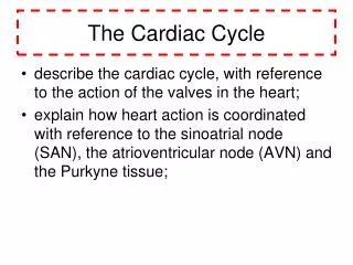
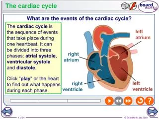
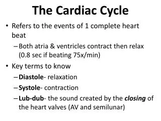
Heart Tones • S1—closure of the AV valves (tricuspid and mitral valve) • --beginning of systole, heard best at the apex, lower in pitch and a bit longer than S2 • S2– closure of the semilunar valves (aortic and pulmonic) at end of systole, best heard in aortic and pulmonic areas, higher pitch and shorter than S1.
Heart tones • S3—During diastole, passive blood filling ventricles, distending walls and causing vibration. Low pitched , heard best at apex or LSB with bell of stethoscope. • S4—During late diastole, caused from vibration in the valves, papillae, and ventricular walls.
Terms of heart tones • “Splitting”—failure of mitral & tricuspid or aortic & pulmonic valves to close simultaneously. • “clicks”—sometimes heard with prolapse or stenosis of valves; often accompanies murmurs, and should always be considered pathologic.
Extra Heart Sounds • Friction rub-caused by pericardial inflammation as in with pericarditis or pericardial effusion; can hear after an MI. --harsh grating sound; not always heard consistently. Sounds like the sound made when walking on snow that has frozen over.
Heart Murmurs • Grade I- very faint, not heard in all positions • Grade II- soft but easily heard • Grade III- moderately loud • Grade IV- loud and may be associated with a thrill • Grade V- very loud, heard with stethoscope barely on the chest, thrill present • Grade VI- may be heard with stethoscope off chest; associated with a thrill.
Heart Murmurs • Describe: • Grade- I,II,III,IV,V,VI • Frequency- (pitch)-harsh,rumbling,blowing,musical • Timing- midsystolic,holosystolic, early systolic, late systolic, early diastolic, mid-diastolic, late diastolic • Shape- crescendo,decrescendo,plateau • Location- on chest where murmur is loudest • Radiation- to neck, axillae
(Example) Aortic stenosis murmur • Grade III/VI, harsh, crescendo-decrescendo, midsystolic, murmur heard best at 2nd ICS, right sternal border, with radiation to the neck.
Heart Murmurs If you hear a murmur: Assess: • Has pt has ever been told they have a murmur? • Any diagnostic testing ever done (echo)? • Any signs or symptoms such as SOB, palpitations, syncope, pre-syncope, CP, edema?
Words of Wisdom: • If you think you hear it, you probably do. • If it’s a murmur, get an echo
Atrial Fibrillation Michelle Maloney MSN, APRN, ANP-BC
Atrial Fibrillation • The most common sustained cardiac rhythm abnormality • Increases in prevalence with age, rates of 5-10% in those over the age of 80 • More common in men
Classifications • Paroxysmal AF refers to patients with spontaneous termination of the arrhythmia within 7 days of onset. Most patients spontaneously going in and out of AF on their own fall into this category. • Persistent AF refers to patients with sustained arrhythmia beyond 7 days. Most patients in this category require the use of a therapeutic intervention to restore normal sinus rhythm. • Permanent AF refers to patients in which efforts to restore normal sinus rhythm have either failed or been forgone.
Causes of atrial fibrillation • Surgical procedure • Pulmonary disease (COPD, PE) • Thyroid disease (hypo- and hyper-) • Hypertensive heart disease • Acute or chronic alcohol use • Myocardial infarction • Obstructive sleep apnea • Valvular heart disease • Sepsis/pneumonia
Symptoms of atrial fibrillation • Palpitations • Nonspecific complaints: dyspnea, fatigue, lightheadedness, dizziness • Symptoms may be more pronounced in patients with LV systolic dysfunction (loss of atrial contraction and RVR decrease cardiac output and increase congestive symptoms) • PATIENTS MAY BE ASYMPTOMATIC!
Management of Atrial Fibrillation • Three fundamental aspects to management of atrial fib: • Rate control • Prevention of thromboembolism • Rhythm control (may not be necessary in all patients, primarily directed by symptoms associated with AF) • Unstable patients with symptoms of chest pain, hypotension, pulmonary edema may require emergent cardioversion
Rate Control • Aim for HR slower than 80 at rest and slower than 110 with moderate exercise • Utilize drugs that prolong refractory period of the AV node: • Beta blockers (metoprolol, atenolol, nadolol, carvedilol) • Calcium channel blockers (diltiazem, verapamil) • Digoxin (typically an add on with above meds) • Amiodarone
Precautions with medications • Beta blockers: hypotension, contraindicated in pts with bronchospasm • Calcium channel blockers: hypotension, may worsen heart failure in decompensated pts • Digoxin: narrow therapeutic window, effective at slowing HR at rest but not during activity • Amiodarone: long term use with many side effects (thyroid, pulmonary fibrosis, visual changes)
Several risk classification schemes to risk-stratify patients into high-risk or low-risk for stroke associated with atrial fibrillation -Most useful is CHADS2 (five features) -Another is the CHADS2-VASC2 (same 5 features plus additional features)
CHADS2 Score 0: Low risk, no anticoagulation or aspirin only Score 1: Moderate risk. Aspirin daily or Warfarin, INR 2.0-3.0, or new anticoagulant Score 2: Moderate or High risk. Warfarin, INR 2.0-3.0, or new anticoagulant
CHADS2-VASc Score 0: Low risk. No antithrombotic or aspirin daily. Score 1: Moderate risk. Oral anticoagulation or aspirin. Warfarin INR 2.0-3.0 or new anticoagulant. Score 2 or >: High risk. Oral anticoagulation. Warfarin INR 2.0-3.0 or new anticoagulant.
Anticoagulation Warfarin Newer Oral Anticoagulants (Rivaroxaban, Dabigatron, Apixaban) PROS: No lab testing required Standard dosing (with renal dose adjustment) Quicker onset CONS: Expensive No antidote or reversing agent PROS: • Inexpensive • Reversible with administration of Vitamin K CONS: • Slow onset of action • Narrow therapeutic index requires regular lab testing • Numerous drug-drug and drug-food interactions
Newer Anticoagulants Dabigatran (Pradaxa) – 150mg PO BID -BID dosing -10% of patients experience dyspepsia -Renal dosing Rivaroxaban (Xarelto) —20mg PO daily -Once daily dosing -Renal dosing Apixaban (Eliquis) – 5 mg PO BID -BID dosing -Lower dosing if 2 of the following: age >/= 80, body weight </= 60kg, serum creatinine 1.5 or higher **All are contraindicated in patients with mechanical prosthetic heart valves**
Rhythm Control • Several clinical trials have shown no overall difference in mortality in rate-control versus rhythm-control (both strategies including anticoagulation therapies) • Rhythm control should be individualized decision based on symptoms associated with AF, patient preferences, and response to treatment
Pharmacologic Rhythm Control Propafenone (Rhythmol), Flecainide, Sotolol • For recurrent paroxysmal atrial fibrillation • For patients with no or minimal heart disease • Contraindicated in patients with CAD or significant LVH • Sotolol used with caution in those with renal insufficiency
Pharmacologic Rhythm Control (cont) Amiodarone • Can be used in patients with heart failure • Long term use based on risk-to-benefit ratio because of known side effects (vision, thyroid, pulmonary) Dronedarone (Multaq) (an amiodarone analog) • Fewer side effects than amiodarone. For paroxysmal atrial fibrillation. • Contraindicated in patients with severe heart failure. • Should not be used for patients with permanent atrial fibrillation (increased risk of cardiovascular death)
Cardioversion • Immediate cardioversion can performed if duration of the atrial fibrillation is known to be less than 48 hours • Conversion success rate higher for patients in AF for shorter duration • Can be done with pharmacologic agents (amiodarone, ibutilide) or direct-current electrical cardioversion
-For atrial fibrillation of 48 hours’ duration or longer (or when duration is unknown), anticoagulation is recommended for at least 3 weeks before and 4 weeks after cardioversion, regardless of method used for cardioversion. -Anticoagulation should be continued after cardioversion until sinus rhythm has been maintained for at least 4 weeks
TEE (Transesophageal Echocardiogram) • As an alternative to anticoagulation prior to cardioversion (or for patients who cannot complete the 3 full weeks of anticoagulation prior to procedure), a transesophageal ECHO can be performed to exclude presence of a left atrial thrombus • Even in individuals with no left atrial thrombus, anticoagulation after cardioversion is still necessary
Procedure-Based Treatments for Atrial Fibrillation • Catheter Ablation: ideal candidate is a patient with paroxysmal AF in the absence of structural heart disease. Primary focus is electrical isolation of the pulmonary veins • Surgical Ablation: the Maze procedure. Involves incisions to atrial tissue to interrupt abnormal reentry circuits. Reserved for patients undergoing cardiac surgery.