Download
1 / 31
320 likes | 513 Views
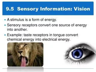
9.5 Sensory Information: Vision. A stimulus is a form of energy. Sensory receptors convert one source of energy into another. Example: taste receptors in tongue convert chemical energy into electrical energy. Sensory Receptors. Highly modified ends of sensory neurons

E N D
9.5 Sensory Information: Vision • A stimulus is a form of energy. • Sensory receptors convert one source of energy into another. • Example: taste receptors in tongue convert chemical energy into electrical energy.
Sensory Receptors • Highly modified ends of sensory neurons • Often different sensory receptors are grouped in specialized sensory organs • Amplifies stimuli * See table 1, pp. 438
Sensory Receptors • Different sensory receptors are frequently grouped together with connective tissue and sensory organs to form structures such as the eye and ear. • A network of touch, high-temperature and low-temperature receptors are found throughout the skin. The brain registers and interprets the sensation.
Sensory Adaptation • When a receptor becomes accustomed to the stimulus, and the neuron ceases to fire, even though the stimulus is still present Example: Receptors are stimulated in your skin when you take on or off your clothes, but sensory information assuring you that your clothes are still on your body is not needed.
Anatomy of the Eye • The eye has three separate layers: the sclera, the choroids layer and the retina.
Eye Anatomy • Sclera: the outermost layer that protects the eye and maintains the shape. It is white and fibrous, but clear in the front where there is a bulging cornea. • Cornea: acts as the window to the eye by bending light toward the pupil. The cornea needs nutrients and oxygen, but obviously does not have blood vessels, so it gets the oxygen from dissolved gases in tears and the nutrients from the aqueous humour (transparent fluid in a chamber behind the cornea)
Eye Anatomy • The middle layer is the choroid layer that contains blood vessels and dark pigments that absorb light to stop reflection. Near the front of the choroid layer is the iris. • The iris is an opaque disk of tissue surrounding the pupil that regulates the amount of light that can enter the eye. • It is made of circular muscle, and acts as a diaphragm
Eye Anatomy • The lens is found just behind the iris and it focuses the image onto the retina. • It has ciliary muscles attached to ligaments that are suspended from the dorsal and ventral ends of the lens that alter the shape of the lens. • A large chamber behind the lens called the vitreous humour, contains a cloudy material that maintains the shape of the eyeball
Eye Anatomy • The innermost layer is called the retina. It has three layers of cells within it: • The light-sensitive cell layer is directly next to the choroid layer. Two types of light-sensitive cells: • Rods: respond to low-intensity light and detect black and white. • Cones: respond to high-intensity light and colour. • The bipolar layer is next and takes the nerve message from the rods and cones, and sends it to the optic nerve cells. • The optic nerve cells are the last layer in the retina and they carry the impulse to the CNS
Eye Anatomy • The centre of the retina is a tiny depression called the fovea centralis. It is the most sensitive part of the eye where the cones are closely packed together. • There are no rods or cones where the optic nerve comes in contact with the retina, so there is a blind spot that results.
Eye Structure The Sense of Sight - Learning Activity Anatomy of the Eye
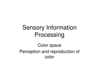
Contemporary Theories of Vision Two complementary theories: Newton: Particles of light (photons), travel in waves of various lengths Huygens: Light enters the eye as it is reflected or transmitted from objects
An old school camera vs. your eyes Camera (eye part in brackets): • Diaphragm opens and closes to let light in and block light out • Image of camera is focused on a film
Afterimages • Positive and negative • Positive occurs after you look into a bright light and then close your eyes • Negative occurs after you look at a bright coloured light for a long period of time
Focusing the Image • As light enters the eye, it is bent toward the pupil by the cornea. • Light bending is called refraction. • The cornea directs the light toward the lens, and it bends further. • Because the lens is biconvex () the light is bent toward a focal point and an inverted image is projected onto the retina. • When we view close objects, the ciliary muscles that control the shape of the lens contracts, and the tension on the ligaments decrease, making the lens thicker. • When we view objects far away, the relaxation of the ciliary muscles causes the tension on the ligaments to increase, making the lens thinner. • These adjustments made by the lens and pupil are referred to as accommodation. • Objects that are 6m away do not need any accommodation. • As we age, the lens looses its flexibility, and we usually have a hard time reading up close. Parasympathetic response
In Summary • Light enters the eye, and is bent towards the pupil by the cornea • The dense cornea bends the light by slowing it down refraction • The biconvex lens in the cornea bends light further inwards, because it is thicker in the middle than at the edges
Summary • The image is inverted on the retina • Ciliary muscles control the shape of the lens, while ligaments maintain a constant tension Viewing Close Objects: • Ciliary muscles contract, ligament tension decreases • Lens becomes thicker…additional light bending • Pupil constricts Viewing Far Away Objects: • Ciliary muscles relax…increasing ligament tension • Lens becomes thinner • Pupil dilates Visual Pathways
Accomodation • Adapting the eyes to objects near and far away • By age 40 your near point accommodation has diminished…trouble reading
Glaucoma • Buildup of aqueous humour in the anterior chamber of the eye • Drainage ducts are blocked • Without constant nutrients and oxygen, neurons die
Cataracts • Lens becomes opaque preventing some light from passing through • Traditionally treated by removing the lens, and giving the patient strong eye glasses
Astigmatism • The lens and cornea are irregularly shaped • Creates a non-distinct focal point
Nearsightedness (myopia) • Eyeball is too long • Lens cannot flatten enough to project the image on the retina, so the image is focused in front of the retina • Allows focus on close objects, but difficulty with distance vision
Farsightedness (hyperopia) • Eyeball is too short • Distant images are brought into focus behind the retina • May focus on objects in the distance, but will have trouble seeing near objects