Download
1 / 27
400 likes | 3.05k Views
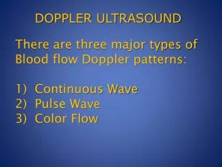
There are three major types of Blood flow Doppler patterns: 1) Continuous Wave 2) Pulse Wave 3) Color Flow. DOPPLER ULTRASOUND. What can We Measure with Doppler Ultrasound?. Velocities: Most calculations start with this parameter Direction of Blood Flow Pressure Gradients.

E N D
There are three major types of Blood flow Doppler patterns:1) Continuous Wave2) Pulse Wave3) Color Flow DOPPLER ULTRASOUND
What can We Measure with Doppler Ultrasound? • Velocities: Most calculations start with this parameter • Direction of Blood Flow • Pressure Gradients
Beam alignment is crucial for accurate velocity measurements V = velocity of blood flow C = speed of sound in blood FT = transducer frequency FS = backscatter frequency Cos ϴ = cosine of angle between US beam & blood flow
As the angle of the incident beam and the direction of blood increases the measured velocity decreases and the error increases DOPPLER BEAM MEASURED BLOOD FLOW VELOCITY VECTOR WHEN BEAM IS NOT PARALELL BLOOD FLOW VELOCITY VECTOR
Effects of Intercept Angle in Velocity Measurements can not be ignored beyond 20 degrees
Color Flow Principle is similar to PW with multiple sector scans
SPECIFIC APPLICATION OF DOPPLER MEASUREMENTS • Pulsed Wave Doppler • Stroke Volume Measurement • Continuous Wave Doppler • Peak velocity measurement • Color Doppler • Blood Volume flow & direction
Pulsed Wave Doppler • Measures the blood velocity at a specific location or sample volume • Exact localization of moving objects • Limited maximum velocity measurement because of Nyquist Limit (more on this later)
Pulsed Wave Doppler from transmittal flow to evaluate diastolic function
Continuous Wave Doppler • Measures the Maximum Velocity in a specific direction ( vs location for PW) • Essentially no limit in the maximum velocity measurement (no Nyquist limit) • The site of the maximum velocity cannot be localized
Continuous Wave Doppler MR JET VELOCITY PROFILE DURING SYSTOLE, NOTE DIRECTION AND MAX VELOCITY
Color Doppler • Multiple sampling with pulsed wave Doppler along the 2D scan lines. • The quantity and direction of velocity are measured in every point. • The intensity of the signal represents number of moving cells • Movement away from the transducer is blue and movement toward the transducer is red.
Blood Velocity Curves • Can be measured any where there is flow and an Acoustic Window • Basic calculus is used to analyze the velocity curves to obtain the Stroke Volume (SV) and Cardiac Output
CONTINUOUS DOPPLER WAVES CAN BE USED TO MEASURE:STROKE VOLUMECARDIAC OUTPUT
STROKE VOLUME & CARDIAC OUTPUT INTERGRAL OF BLOOD VELOCITY DISTANCE TRAVELED BY BLOOD DISTANCE TRAVELED BY BLOOD AREA OF FLOW VOLUME OF BLOOD (STROKE VOLUME)
Stroke Volume Measurement thru LV out flow tract (LVOT) • SV = CSALVOT x VTILVOT SV = Stroke Volume CSA = Cross Sectional Area VTI = Velocity Time Integral LVOT = Left Ventricular Outflow Track
Continuous Wave Doppler for Pressure Gradient • Pressure Gradient = 4 x V2 • Pressure Gradient = 4 x V21 – V22 • Measuring maximum gradient through the narrowest area of the flow • Requires measuring the maximum velocity thru the area of flow with CW
SUMMARY • Doppler measurements have numerous applications in echocardiography. • We can also measure the velocity of intra-cardiac structures (myocardium or valves) and assess their function or dysfunction.
SUMMARY • Understanding Echocardiography Physics will enhance our understanding of: • The physical principles that affect our measurements and images • Knowing when to believe or doubt our results • The strengths and limitations of the technology