Guillain-Barre Syndrome
640 likes | 1.88k Views
Guillain-Barre Syndrome. William Woodfin MD. K.F. 40 y.o. r/h woman. 3/17 Nausea, diarrhea & severe myalgias Son dxed c rotavirus 1 wk. Previously 4/21 “Creepy-crawlies” legs>arms 4/25 Weakness legs progressing 4/26 Handwriting looks like “hen scratch”. K.F. 40 y.o. woman.

Guillain-Barre Syndrome
E N D
Presentation Transcript
Guillain-Barre Syndrome William Woodfin MD
K.F. 40 y.o. r/h woman 3/17 Nausea, diarrhea & severe myalgias Son dxed c rotavirus 1 wk. Previously 4/21 “Creepy-crawlies” legs>arms 4/25 Weakness legs progressing 4/26 Handwriting looks like “hen scratch”
K.F. 40 y.o. woman 4/28 Admitted to outside hospital. L.P. wnl EMG positive waves in some leg muscles NCVs absent H-reflexes F responses & motor latencies wnl
K.F. 40 y.o. woman 4/29 Transferred to PHD Hx.: diabetic x 10 yrs. hypothyroid- treatedx yrs. no sphincter disrubance aching pain low back & buttocks mild postural light headedness no SOB or palpatations
Exam BP 150/90 P 80 Wt. 250 lbs. Mild weakness neck flexors 4/5 biceps, grip & interossei- symmetric 2/5 iliopsoas & quadriceps 3/5 hamstrings & adductors 4/5 abductors 4/5 ankles & toes- extensors & flexors
Exam Sensory- intact DTRs- biceps, BR, knees are trace c reinforcement. Triceps & ankles unobtainable Plantars- flexor F to N- intact Gait- not testable
Lab H/H 10.3/33.5 c microcytic indices A1c Hgb 10.1 TSH 0.97 LDL 182 Serum immunofixation- wnl. No IgA def. FVCs- consistently 4+ liters
MRI LS spine s & c contrast- no nerve root enhancement
Course in hospital Treated c IVIG 0.4 gms/kgm daily x 5 Strength fluctuated only mildly Blood sugars ok in AM, high in afternoons Repeated NCVs show mild dispersion of F waves Transferred back to referring hospital 5/6
Telephone FU Ambulating fairly well c walker. Strength clearly improving. Still bothered by “creepy-crawlies”
What is the GBS? • Due to the breadth of clinical presentation it is of limited help to try to define rigid diagnostic criteria. • Thomas Munsat 1965: “…The GBS is easy to diagnose but difficult to define
1. Paresthesiaes usually hearld the disease 2. Fairly symmetric weakness in the legs, later the arms and, often, respiratory and facial muscles 3. Dimunition and loss of the DTRs 4. Albuminocytologic dissociation 5. Recovery over weeks to months The typical illness evolves over weeks usually following an infectious disease and involves:
Waldrop 1834 Olliver 1837 Landry 1859 Graves 1884 Ross & Bury 1893 Brussel’s Conf. 1937 Haymaker & Kernohan 1949 Waksman & Adams 1955 Miller Fisher 1956 Asbury, Aranson & Adams 1969 History Guillain, Barre & Strohl 1916-1920
Note sur la paralysie ascendante aigue 1859 • March 16- a febrile illness • May 11- mild sensory symptoms in the fingers and toes • June 13- knees buckle • June 16- unable to walk • Subsequent respiratory failure and death. • Autopsy unrevealing. Peripheral nerves probably not examined
Late 19th century • Westphal 1876- “Landry’s Ascending Paralysis” • Graves 1884- localized neurologic disease to the peripheral nerves, “the nervous cords” • Ross & Bury 1893- 90 cases. A disease of the peripheral nerves • Numerous reports emphasizing various aspects of the disease with most authors crediting Landry
Guillain, Barre & Strohl 1916Revue Neurologique • Two soldiers in Amiens developing paralysis and loss of DTRs. • A new diagnostic feature: albuminocytologic dissociation in the CSF • No mention of Landry
Foundations • Quincke- CSF observations 25 years earlier • Siccard & Foix- “albuminocytologic dissociation” in Pott’s disease Late 19th century: examination of the reflexes had become a part of the neurologic exam with appreciated as a sign of neuropathy based on observations in tabes dorsalis areflexia
Haymaker & Kernohan 1949 • Landmark in pathological description c 50 fatal cases & detailed review of clinical findings • Emphasized prominent damage to proximal nerves often at junction of ventral & dorsal roots. Little study of more distal nerves • Unified findings of Landry & Guillain, Barre & Strohl
Waksman & Adams 1955 • Experimental Allergic Neuritis • First animal model of a noninfectious inflammatory neuritis • Rabbit nerve and Freund’s adjuvant injected intradermally • Target of activated T cells uncertain
Asbury, Aranson & Adams 1969 • 19 pts. All with well developed mononuclear infiltrates in spinal roots and nerves within days of clinical onset • Pathological hallmark: perivascular mononuclear inflammatory infiltrates to adjacent to the areas of demyelination
Overview of Adaptive Immunity • Lymphocytes: “command & control,” identify antigen components, respond specifically, mobilize other elements and direct the attack c memory for each antigenic assault • Antibodies: specialized immunoglobulin molecules directly neutralize and remove antigen
T lymphocytes • CD8- recognize epitopes paired c MHC-I • CD4- activate and control the immune response • Scavenger cells break down antigen into small peptide fragments (T cell epitopes), MHC-II epitope complexes are expressed on the surface & the scavenger become an APC which docks on a CD4 c a compatible TCR. CD4 proliferates releasing cytokines.
Antibodies • Cytokines activate other lymphocytes including B cells that differentiate into plasma cells and serve as immunoglobulin factories. • Abs are Ig molecules that recognize, bind, neutralize and opsonize Ag for phagocytosis. They activate complement(membrane attack complex) & induce target cells to activate the inflammatory response
Self-tolerance • The process of self recognition • T & B cells learn self tolerance during maturation • Autoimmunity occurs when the mechanisms of self protection are defective
Mechanisms of Autoimmunity • Molecular mimicry- microbe cell surface Ag resembles self protein. Damage results from “friendly fire” The inciting Ag is usually unidentified & may not exist as a single stimulus. • Excessive cytokine release due to profound immune stimulus may awaken self tolerant T cells or may cause expression of MHC complexes. • Self Ags bound to drugs may lose tolerated status
Antecedent Events: Infectious • Viral: Influenza, Coxsackie, EBV, Herpes, HIV, Hepatitis, CMV, WNV Bacterial: Campylobacter jejuni, Mycoplasma, E. coli Parasitic: Malaria, Toxoplasmosis
Antecedent Events: Systemic disease • Hodgkins • CLL • Hyperthyroidism • Sarcoidosis • Collagen Vascular d. • Renal d.
Other antecedent events • Surgery • Immunization • Pregnancy • Envenomization • Bone marrow transplantation • Drug ingestion
Features of AIDP • 2/3s have identifiable preceding event • 50% begin with paresthesias followed by weakness in legs; 10% begin with arm weakness; rarely begins in face • Ophthalmoplegia: partial 15%, total 5% • Autonomic dysfunction in 65%, arrhythmias, hypotension,urinary retention in 10-15%, pupillary inequality
AIDP • Progresses for days to 4 weeks • 15% with severe disability • Mortality 3-5% • CSF: protein may be normal early, elevated in 90% by clinical nadir, cells< 10 in 95%, >50 suggests HIV • EDX: prolonged F & distal motor latencies, conduction block 30-40% in routine studies
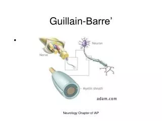
AIDP • Pathology: immune attack directed at schwann cell plasmalemma esp. at nerve roots with IgG & complement deposits preceding demyelination
CIDP • Evolves over months • Fluctuates • Respiratory failure, dysautonomia, facial weakness, ophthalmoplegia- all are rare • CSF protein often highly elevated • Marked slowing of motor nerve conduction • Steroid responsive
Features of AMSAN • Commonly preceded by diarrhea esp. c. jejuni • Abrupt onset of weakness c rapid progression to quadriplegia & respiratory insufficiency • Other features as c AIDP • Longer recovery, more residual & mortality 10-15%
AMSAN • CSF as in AIDP • EDX: no response in some motor nerves, decreased amplitude of the CMAPs, fibrillations on needle study, absent SNAPs • Immune attack directed at axon plasmalemma at nodes of Ranvier. Wallerian degeneration
Features of AMAN • Often preceded by diarrhea affecting younger population in China. Sporadic in USA • Prognosis similair to AIDP • Mortality <5% • EDX: reduced CMAPs c normal F & distal motor latencies and sensory studies. Fibrillations in 2-3 weeks
AMAN • Pathology: again axonal plasmalemma at nodes of Ranvier sometimes limited to physiologic dysfunction c nodal lengthening. May go on to extension through axonal basal lamina. Most axons recover s Wallerian degeneration
Miller Fisher Syndrome • Ophthalmoplegia, Ataxia, Areflexia • May be heterogonous: 1. Related to other patterns of GBS 2. Related to brainstem encephalitis, Bickerstaff 1952 3. CNS demyelination in association with GBS
Miller Fisher Syndrome • 95% have serum IgG Ab to ganglioside GQ1b • Studies show preferential location of anti-GQ1b to cerebellar molecular layer & Cranial Nerves 3,4 & 6 • May act at N-M junction depleting acetylcholine from nerve terminals
Acute Panautonomic Neuropathy • Manifests over 1-2 weeks but may be of subacute onset • Frequent preceding infection • DTRs lost in 1/3, distal sensory changes 1/4 • Albuminocytologic dissociation • EDX: NCVs usually normal • Recovery is gradual and incomplete
Differential Diagnosis • Consider the possibility of an upper motor neuron lesion • Other considerations are rare. Diphtheritic neuritis & poliomyelitis belong more to the history section of this presentation. A new possibility is West Nile Virus.
Differential • N-M: MG, LES, Antibiotics • Toxic: Cigutera (ciguatoxin), Pufferfish (tetrodotoxin), Shellfish (saxitioxin), Botulism, Tick paralysis (Lone Star tick, Gulf Coast tick), Glue sniffing, Buckthorn • Mononeuritis multiplex assoc. c Wegner’s. PAN, SLE, RA, Sjogren’s, Cryoglobulinemia etc.
Differential • Metabolic: Periodic paralyses, Hypokalemia, Hypermagnesemia, Hypophoshatemia c parenteral hyperailimentation, Thyrotoxicosis, ICU myoneuropathy (CIP) • Heavy metal: Lead, Arsenic, Thallium, Barium c hypokalemia
Differential: Miller Fisher Syn. • Multiple sclerosis • Encephalitis • Posterior circulation ischemia or infarct • Other: Botulism, MG, Tick
Treatment Respiratory failure Autonomic dysfunction DVT & PE Pain Positioning & Skin care Physical therapy Nutrition