Download
1 / 76
1.5k likes | 3.78k Views
Adolescent Idiopathic Scoliosis. Sasha Carsen Dr. Jarvis Dec 2, 2010. Outline. Background Definition Epidemiology/Demographics Pathoetiology Clinical Evaluation History Physical Exam Management Non-Operative Operative (& indications) Practice/OITE Q’s. Normal Spinal Curves.

E N D
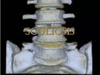
Adolescent Idiopathic Scoliosis Sasha Carsen Dr. Jarvis Dec 2, 2010
Outline • Background • Definition • Epidemiology/Demographics • Pathoetiology • Clinical Evaluation • History • Physical Exam • Management • Non-Operative • Operative (& indications) • Practice/OITE Q’s
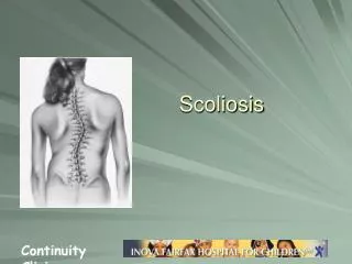
Normal Spinal Curves • Frontal/Coronal plane • Straight • Thoracic Kyphosis • 20-40 deg • Lumbar Lordosis • 30-60 deg
Types of Scoliosis • Congenital • Neuromuscular • CP, Myelomeningocele, etc. • Syndromic • Secondary to Tumor, Trauma, Paralytic, etc. • Idiopathic • Most Common • Scoliosis defined as Cobb > 10 deg
Idiopathic Scoliosis DEFINITION Structural lateral and rotational deformity of the spine for which no cause can be established. “Idiopathic, from the Latin meaning we're idiots 'cause we can't figure out what's causing it.” Dr. Gregory House, 2005 (referring to Idiopathic T-cell deficiency, not AIS)
Idiopathic Scoliosis Types: • Infantile (0-3) - <1% • Juvenile (4-9) - 10.5-12% • Adolescent (10 to maturity) – 89% • Dickson: Early 0-5 vs Late 5-Maturity
Etiology • Multiple theories have been hypothesized • Genetic • Not a single locus, likely polygenic • First noticed/reported in ‘60’s by Wynne-Davies, found >11% rate in 1st degree relatives • Later twin studies showed 73% in Monozygotic twins, and 36% in Dizygotic (Kesling, Reinker)
Etiology • Many other theories • Melatonin: Doubousset shown 35% decr in melatonin levels • Dec Type II (fast twitch in paraspinal) • Platelet & calmodulin (melatonin binds (inhibits)) • Genetics and Biomechanics appear to be most important • Bipedal stance a prerequisite. • Bipedal rats (pinealectomized) will get AIS • Scoliosis surgery/bracing in rats very challenging
Genetics • Genetic Test now available that may help predict future progression/severity of scoliosis. • Only useful if strong positive or neg • Evidence may have bias • $$$ • The future? • Axial Biotech (DuPuy Spine) Ogilvie, “The search for idiopathic Scoliosis genes”, Spine, 2006. Ogilvie, “Adolescent Idiopathic Scoliosis and Genetic Testing”, Current Opinion Pediatrics, 2010.
Demographics • Prevalence • 1.5-3% Adolescent Pop have curve >10 deg • 90% of these will not require Tx • 0.15-0.3% curve>25-30 • F>M • Up to 10:1
Clinical Presentation • Asymptomatic • Detected by school screening, pediatrician, or a family member. • School Screening: Controversial • 2004 US Preventive Health Task Force recommended against school screening • 2008 SRS Recommended judicious screening • If presents with back pain must R/O other pathology
History • When was the curve first noticed? • How has it changed since first noticed? • Any associated symptoms, especially neurological, bowel, or bladder changes? • AIS is not a painful condition • Constitutional symptoms suggest other cause • FH of scoliosis?
History • What level of skeletal maturity is the patient at? • Maximal progression occurs during maximal/peak growth velocity • Maximal growth velocity occurs prior to menarche • Females continue spine growth for approximately 1.5-2 years after menarche • Risser stage and menarche are key • Other signs such as tanner staging and bone age determinations (tri-radiate cartilage, etc.) can also be used
Physical Exam • Observation • Maturity • Curve location and magnitude • Balance of shoulders and pelvis, asymmetry • Leg lengths for inequality • LLD can cause apparent scoliosis/rotation • Soft tissue fold asymmetry • Plumb Line – Occiput centred over Sacrum
Physical Exam • Forward bending • Assesses extent of structural rotation • Rib prominence on side of convexity • Legs together, hands together, bending at waist with head down • Look from behind patient • Postural scoliosis would have no rotation • Scoliometer >7deg sensitive for 20 deg • 5-7 deg = 15-20
Neuro Exam • Neurological Exam • AIS normally has no abnormal findings • Key part of any exam • Includes motor, sensory and reflex examination, clonus, UMN • Abnormal abdominal reflex may reflect intra-spinal pathology Abdo Reflex Stroke the abdomen lightly on each side in an inward direction above and below the umbilicus.Contraction of the abdominal muscles and deviation of the umbilicus towards the stimulus.
Syrinx • In classical mythology, Syrinx was a nymph and a follower of Artemis, known for her chastity. Pursued by the amorous Greek god Pan, she ran to the river's edge and asked for assistance from the river nymphs. In answer, she was transformed into hollow water reeds that made a haunting sound when the god's frustrated breath blew across them. Pan cut the reeds to fashion the first set of pipes, which were thenceforth known as syrinx.[1] The word syringe was derived from this word.
Syrinx • A syrinx is a fluid-filled cavity within the spinal cord (syringomyelia) or brain stem (syringobulbia). Predisposing factors include craniocervical junction abnormalities, spinal cord trauma, and spinal cord tumors. • Treatment includes correction of the cause and surgical procedures to drain the syrinx or otherwise open CSF flow.
Investigations • AP and LAT radiographs first step in diagnosis • Standing film • One long cassette • Assessment of curve type , magnitude, and and bony indicators of maturity
Investigations • XR assessment • Cobb angle: angle subtended by vertebrae at ends of curves • >10 deg • Risser sign: extent of iliac apophysis calcification • 0-5 (F spinal growth stops 4)
Further Imaging • XR • Bending Films • Traction Films • Degree of flexible vs. fixed deformity • MRI • Left thoracic curve • Neurological abnormalities • Noted structural abnormalities on X-ray • Excessive kyphosis • Rapid progression • Juvenile onset • Identifiable syndrome • Male gender
Classification • Based on curve and spine characteristics • Helps to guide Tx/Surgical approach • King • Based around Thoracic curve, designed for use of Harrington and like systems (before widespread pedicle screws) • Poor Kappa • Lenke • Thoracic and Lumbar • Include Lumbar and Kyphosis modifiers • Guides Tx in the Pedicle screw age
CSVL = Central Sacral Vertebral Line King • Lumbar > Thoracic, • Thoracic > lumber which crosses the CSVL • Similar to 2 but the lumbar curve doesn’t cross the CSVL • Single long thoracic curve • Double thoracic curve
King 3 King 4 KING 3 KING 4
Lenke • Triad: • Curve type 1-6 • Lumbar modifier (A,B,C) (central sacral vert line) • A – bet pedicles (low risk) 41% • B – to disk/body (mod risk) 37% • C – Lat to body (high risk) 22% • Sagital thoracic (-, N, +) • - = hypokyphotic <10 14% • N = nl kyphosis 10-40 75% • + = hyperkyphotic >40 11%
Treatment Treatment based on patient- and spine-specific factors Options • Observation • Bracing • Surgery Electrical stimulation, exercise, chiropractic… no evidence to support. No ‘prescribed’ role.
Observation • Curve < 25 degrees • Repeat clinical exam and x-rays every 4-12 mo depending on age • Patients within the peak/rapid phase of growth • f/u every 4-6 mo • If curves progress > 5 degrees bracing is indicated • Curves > 30 degrees in the skeletally mature patient are monitored every 5 years
Bracing • For curves>25 and <45 degrees • May halt or reduce curve progression • Does not reverse the deformity • Results are dose dependent • Compliance! • Worn up to 23 hours a day or during sleep depending on the type of brace
Bracing Indications • Skeletal immature patient with a curve of 25 to 45 degrees • The deformity must be cosmetically acceptable by the patient • Patient must be compliant • >20hrs/day (except Charleston-type) • Some newer braces have temp/touch sensors • Contraindicated with lordotic or hypokyphotic curves Consider: • TLSO (Boston)- curves below T8 • Milwaukee- above T8
Bracing • Does it work? • Some questions still being asked • RTCs under way • Cochrane Review 2010: “Bracers for idiopathic scoliosis in adolescents” • Currently very poor quality of evidence • Despite lack of prospective RCTs, there is a significant body of literature supporting bracing • Schiller et al. “Brace management in AIS”, CORR, 2010
Surgery • Goals: • Arrest progression • Achieve 3D correction (as possible) • Improve appearance, balance trunk • Keep complications of curves/progression/Tx to a minimum • Surgical Outcomes hotly debated • How to define? Radiographic? Curve arrest? QOL? • Howard et al. (SickKids) “Improvement in quality of life following surgery for Adolescent Idiopathic Scoliosis”, Spine, 2007
Surgery Indications • Thoracic curves >40-50 degrees in a skeletally immature patient • Thoracic curve >50 in a skeletally mature patient • Severe trunk shift and vertebral rotation
Surgery Pre op planning • Adequate clinical and radiological assessment • Intra op neurological monitoring • SSEP and MEP • Wake up test gold standard • Autologous blood transfusion and cell saver • Post op ICU admission
Surgery Procedures Fusion! • ASF • PSF • ASF+PSF • Some discussion in literature of stapling, other novel surgical Tx, these are not the mainstay • (won’t be the exam answer)
Pedicle Screws • Initial hesitance in adoption, concerns about safety & learning curve • Hicks et al. “Complications of pedicle screw fixation in scoliosis surgery: A systematic review”, Spine, 2010. • Over 4500 pedicle screws placed, approx 11% malpositioned • No significant clinical complications • $$
Approach • PSF and instrumentation remains the gold standard for thoracic and double major curves • ASF+PSF is indicated for some curves > 75 degrees and in patients at risk of crankshaft deformity • Large curve, skeletally immature