Effectiveness of Orthoses and Foot Training in Patellofemoral Pain
10 likes | 103 Views
This study investigates the effectiveness of a standardized foot training program combined with foot orthoses in patients with Patellofemoral Pain Syndrome (PFP) who exhibit hyperpronation. Patients underwent supervised exercises weekly for three months. The study showed significant improvement in pain and symptoms in the intervention group compared to controls, with long-lasting effects up to twelve months. The control group offered a standard training regime showed no improvement. Foot training and orthoses could be crucial in managing PFP.

Effectiveness of Orthoses and Foot Training in Patellofemoral Pain
E N D
Presentation Transcript
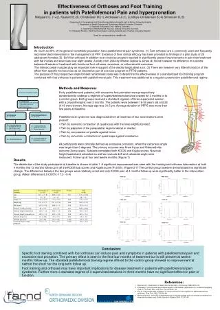
Introduction As much as 25% of the general nonathletic population have patellofemoral pain syndrome. (1) Foot orthoses are a commonly used and frequently recommended intervention in the management of PFP. Evidence of their clinical efficacy has been provided by findings of a pilot study of 20 adolescent females (3). Soft foot orhtoses in addition to an exercise program resulted in significantly greater improvements in pain than treatment with flat insoles and exercises over eight weeks. A study from 2004 by Wiener-Ogilvie & Jones (4) found however no difference in outcome between 8 weeks of treatment with functional foot orthoses, exercises, or orthoses with exercises. The intrinsic pedal muscles play an important role in support of the medial longitudinal arch. (2) There are however very little information of the effect from specific foot exercise as an imperative part of exercise program to PFPS patients. The purpose of this prospective single blinded randomised study was to determine the effectiveness of a standardized foot training program combined with foot orthoses in patients with patellofemoral pain. This treatment was additional to a regular conservative patellofemoral regime. Effectiveness of Orthoses and Foot Trainingin patients with Patellofemoral Pain and hyperpronationMølgaard C. (1+2), Kaalund S.(3), Christensen M.(1), Andreasen J.(1), Lundbye-Christensen S.(4) Simonsen O.(5) 1. Department of Occupational and Physiotherapy Aalborg Hospital, part of Aarhus University Hospital 2. Department of Health Science and Technology, Aalborg University, Denmark3. Kaalunds Orthopedic Clinic, Aalborg, Denmark. 4. Department of Mathematical Sciences, Aalborg University, Denmark. 5. Orthopedic Division, North Denmark Region, Aalborg Hospital, part of Aarhus University HospitalEmail for correspondence: cmm@rn.dk • Methods and Measures: • Forty patellofemoral patients, with excessive foot pronation were prospectively randomised to undergo a regimen of supervised exercise once a week for 3 months or to a control group. Both groups received a standard regimen of three supervised session with a physiotherapist over 3 months. The patients were between 18-58 years old and 28 of 40 were women. Average age was 31.5 yrs. Average duration of PFPS was more than five years at baseline. • Patellofemoral syndrome was diagnosed when at least two of four examinations were present: • Pain by isometric contraction of quadriceps with the knee slightly bended. • Pain by palpation of the parapatellar regions lateral or medial. • Pain by compression of patella against femur. • Pain by concentric contraction of quadriceps against resistance. • All participants were clinically defined as excessive pronators, when the calcaneus angle was larger than 6 degrees. The primary outcome was Knee Injury and Osteoarthritis outcome Score (pain). Subjects completed both KOOS and Kujala scores. Navicula height loaded and unloaded as well as navicula drift and calcaneal angle were measured. Follow up at four and twelve months.(Figure 1) Results The distribution of the study participants at baseline is shown in table 1. A significant improvement was seen with the training and orthoses intervention at both 4 months and 12 months follow up in all the KOOS sub scores and Kujala score (P<0.05). (Figure 2-7) The control group however demonstrated no significant change. The differences between the two groups were relatively small and only KOOS pain at 4 months follow up were significantly better in the intervention group. (Mean difference 8.9 CI95%:-17.3- -0.4) Conclusion: Specific foot training combined with foot orthoses can reduce pain and symptoms in patients with patellofemoral pain and excessive foot pronation. The primary effect is seen in the first four months of treatment but is still present at twelve months follow-up. The standard patellofemoral training regime offered to the control group showed no improvement at neither the short nor the long term follow up. Foot training and orthoses may have important implications for disease treatment in patients with patellofemoral pain syndrome. Further more a standard regime of 3 supervised sessions in three months have no significant effect on pain or function. • References: • Merchant AC. Classification of patellofemoral disorders. Arthroscopy.1988;4:235-240. • Fiolkowskj P. Intrinsic pedal musculature support of the medial longitudinal arch: an electromyography study. J Foot Ankle Surg. 2003 Nov-Dec;42(6):327-33. • Eng JJ, Pierrynowski MR: Evaluation of soft foot orthotics in the treatment of patellofemoral pain syndrome. Phys Ther 1993, 73(2):62-68. • Wiener-Ogilvie S, Jones RB: A randomised trial of exercise therapy and foot orthoses as treatment for knee pain in primary care. British Journal of Podiatry 2004, 7(2):43-49.