Download

1 / 29
290 likes | 310 Views
Learn about temporary pacemakers, pacer wires, settings, pacer modes, and potential complications in cardiac surgery situations with this comprehensive guide.

E N D
EKGs and Pacemakers Cooper University Hospital School of Perfusion 2015 Michael F. Hancock, CCP
Temporary Pacemakers • Provides electrical impulses to the heart to induce depolarization of myocardium • Often used in CT surgery
Indications for Temporary Pacemakers • Arrhythmias following CT Surgery: • Bradycardia • Tachycardia • Complete Heart Block • Ventricular Standstill • Cardiac Arrest
Pacemaker Capabilities • Sense- will detect intrinsic cardiac conduction activity • Pacemaker will respond by either Inhibiting an impulse or Delivering an impulse • Pace- will deliver an electrical impulse • This energy is delivered to the myocardium via the pacing wires
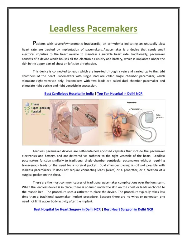
Pacer Wires • Pacemakers rely on pacer wires to deliver the impulse to the myocardium • Types of Pacer Wires: • Epicardial • Transvenous • Transcutaneous Epicardial Pacing Wires
Epicardial Pacing Wires • Surgeon inserts Epicardial Pacing Wires directly into the heart • A Wires- right atrial or Bi-atrial • V Wires- right ventricular or Bi-ventricular
Transvenous Pacers • Routes: • Groin via the Femoral Vein • Neck via a Paceport Swan
Transcutaneous Pads • Go directly on the skin • R-2 Pads • Useful in emergency situations and in minimally invasive procedures • Connected to the Defibrillator
Types of Pacer Wires • Unipolar: (used for Permanent) • One wire into myocardium • One skin wire • Bipolar: (used for Temporary) • Two wires in myocardium • Current flows down one electrode into the myocardium and then back to the pacer via the second wire • Can switch polarities if having issues • Can convert to unipolar with addition of skin electrode
Pacemaker Process • Sense: Pacemaker will first detect intrinsic cardiac conduction activity • Seen on the pacer by “Sense” button flashing • Pacemaker will respond by either Inhibiting an impulse or Delivering an impulse • Pace: Pacemaker will deliver an electrical impulse (“Firing”) • Seen on the pacer by “Pace” button flashing • Capture: When the impulse sent by the pacemaker is received by the myocardium, depolarization will occur • Seen by a pacemaker spike on the EKG
Pacemaker Settings • Rate: • Paces per minute • A/V Output: • Sensitivity measured in mA • Mode: tells pacemaker what to Sense and when to Pace
Pacemaker Process • Selecting a desired Pacing Rate will determine: • PVARP Time: (Post Ventricular Atrial Refractory Period) • If the pacer doesn’t detect activity in this time, it will Fire an impulse and Pace • A-V Interval: • If the pacer doesn’t detect activity in this time, it will Fire an impulse and Pace • Pacemaker Spikes:
Pacemaker Modes Category I: Which chamber is Paced? Category II: Which chamber is Sensed? Category III: What will the pacemaker do if an impulse is Sensed?
Synchronous Pacing • Senses patient’s inherent cardiac activity • Will inhibit or trigger a stimulus as needed • Examples: • AAI, VVI, DDD • Sets a goal for pacing, if the patient doesn’t provide that on their own, then the pacemaker will send the impulse
Asynchronous Pacing • Pacemaker will function at a fixed rate regardless of patient’s conduction • Not able to sense the patient’s underlying rhythm • Examples: AOO, VOO, DOO • Only done if patient has NO intrinsic rhythm!!!
Values at Power ON • DDD • A + V pacing at 80 ppm • mA at 10 each • AV Sense + AV Pace • Pacemaker will look for A+V activity, if none is seen, it will Pace
Emergency Mode • DOO • Asynchronous Pacing • A+V at 80 ppm • Max mA • AV Pacing regardless of patient’s underlying rhythm
Select Pacing Mode • Hit MENU on the pacemaker • Use knob to select desired Mode • Hit SELECT to activate that Mode
Common Settings • AAI • A- Atrium is paced when necessary • A- Atrial activity is being sensed • I = when Atrial activity is sensed the pacer does NOT fire • Ensures Atrial conduction at the given rate
Common Settings • VVI • V- Ventricle is paced when necessary • V- Ventricle activity is being sensed • I = when Ventricular activity is sensed the pacer does NOT fire • Ensures Ventricular conduction at the given rate
Common Settings • DDD • Atrium and Ventricle are paced when necessary (not at same time) • Atrial and Ventricular activity is being sensed by the pacemaker • Dual = • Triggers pacer when Atrial or Ventricular activity is NOT sensed • Inhibits pacer when A or V activity IS sensed
Epicardial Pacing Wires • After around Day 4, the stimulation threshold seems to increase and after Day 5 failure to pace often occurs
Complications ofEpicardial Pacing Wires • Infection • Tamponade • Myocardial damage • Perforation • Disruption of Coronary Anastomosis • Inflammation around site • Increased with higher energy applied
Patient Scenarios • Setting: • Perioperative Cardiac Surgery • Just after Cross Clamp Removal • Rhythm: • Bradycardia • Pacer Mode?
Patient Scenarios • Setting: • Perioperative Cardiac Surgery • Just after CPB • Rhythm: • V. Tachycardia • Pacer Mode?
Patient Scenarios • Setting: • Perioperative Cardiac Surgery • Just after CPB • Rhythm: • A. Fib • Pacer Mode?
Patient Scenarios • Setting: • Perioperative Cardiac Surgery • Just after Cross Clamp Removal • Rhythm: • Asystole • Pacer Mode?