(b)
Supplementary Figure S1. b 3. b 5. b 6. b 7. b 8. (a). b 1. b 2. b 3. b 4. Methyl-K. IgG. G A P S R K ME P D L. R. K P D L R. .V. b 1. b 2. b 3. b 4. IP. Flag-MEF2D. y 7. y 5. y 6. y 3. y 2. y 2. y 1. y 4. y 3. R. K ME2 P D L R. .V. Input. y 2. y 1. y 4.

(b)
E N D
Presentation Transcript
Supplementary Figure S1 b3 b5 b6 b7 b8 (a) b1 b2 b3 b4 Methyl-K IgG G A P S R KME P D L R. K P D L R .V b1 b2 b3 b4 IP Flag-MEF2D y7 y5 y6 y3 y2 y2 y1 y4 y3 R. KME2P D L R .V Input y2 y1 y4 y3 (b) Relative Abundance Relative Abundance Relative Abundance
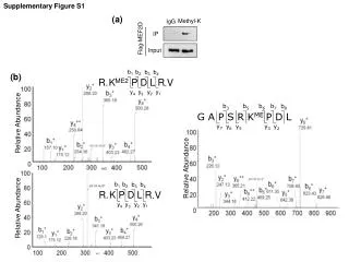
Supplementary Figure S1 b1 b2 b3 b4 R. K P D L R .V (c) b1 b2 b3 b4 y2 y1 y4 y3 R. KME2P D L R .V y2 y1 y4 y3 Relative Abundance Relative Abundance • Supplementary Figure 1.MEF2 is methylated at K267. (a) Transiently transfected Flag-MEF2D in HEK293 cells were immunoprecipitated with normal rabbit IgG or anti-methylated lysine antibody (Methyl-K) and immunoblotted with anti-Flag antibody. (b) Overexpressed Flag-MEF2D in HEK293 cells were immunoprecipitated and analyzed with ESI-LC-MS. Di-methylated (left upper panel), unmodified (left lower panel) and mono-methylated (right panel) MEF2D were detected.(c) Endogenous MEF2D immunoprecipitated from C2C12 cells were analyzed with ESI-LC-MS. Di-methylated (left panel) and unmodified (right panel) MEF2D were detected.
Supplementary Figure S2 (a) (b) (c) (d) MEF2D K267peptide Me0 Me1 IP – + Ionomycin (μg) E14 Diff 0.1 IgG anti-K267me Input IP:anti-K267me IB:anti-MEF2D IP:anti-K267me IB:anti-MEF2D 0.5 1 MEF2D 2 MEF2D IB:anti-MEF2D β-Actin IB: anti-K267me (e) (f) mMEF2A hMEF2A • mMEF2C • hMEF2C • mMEF2D • hMEF2D LGMNSRKPDLRVV LGMNSRKPDLRVV • LGMNNRKPDLRVL • LGMNNRKPDLRVL • LGAPSRKPDLRVI • LGAPSRKPDLRVI H.sapiens M.musculus • P.troglodytes R.norvegicus G.gallus D.rerio LGAPSRKPDLRVI LGAPSRKPDLRVI LGAPSRKPDLRVI LGAPSRKPDLRVI LASNSRKPDLRVI M-ANSRKPDLRVI
Supplementary Figure S2 (g) (h) (i) IP IgG anti-K267me Input WT KR HA-MEF2A Myc-MEF2C WT KR HA-MEF2A IP:anti-K267me IB:anti-HA IP:anti-K267me IB:anti-Myc Myc-MEF2C IB IB: anti-HA IB: anti-Myc HA-MEF2D Supplementary Figure 2. MEF2 is methylated at K267.(a) Anti-methyl K267 (anti-K267me) antibody was tested by dot blot assay using unmodified and chemically mono-methylated K267 containing MEF2D peptides (263-271). (b) Endogenous MEF2D was immunoprecipitated from DO11.10 cells with anti-K267me and immunoblotted with anti-MEF2D antibody. (c) The methylation level of MEF2D K267 in DO11.10 cells treated with Ionomycin (500nM for 3 hours) was analyzed by western blot. (d) mouse embryonic stem cell (E14) were randomly differentiated (Diff) and immunoprecipitated with anti-K267me antibody. (e) Sequence alignment of mouse and human MEF2 family proteins. The methylated lysine residues are highlighted in red. (f) Sequence alignment of MEF2 family proteins between species. The methylated lysine residues are highlighted in red. (g) HA-MEF2A, Myc-MEF2C and HA-MEF2D were overexpressed in HEK293 cells and immunoprecipitated with anti-K267me antibody. (h, i) Transiently expressed HA-MEF2A (h) or Myc-MEF2C (i) wild type (WT) or KR mutant (KR) was immunoprecipitated with anti-K267 methylated MEF2 antibody (anti-K267me) followed by immunoblotting with anti-HA (h) or anti-Myc antibody (i).
Supplementary Figure S3 (a) (b) DM4 GM DM2 G9a * Ezh2 Relative mRNA Expression level * * NTQLGAPSRKPDLRVITSQG MEF2D * * * Myogenin * * * MHC * * Mll2 Ezh2 G9a MHC * Myog SETD6 SETD7 SETDB1 Suv39h1 β-Actin (c) (d) (e) NTQLGAPSRKMEPDLRVITSQG + + – + G9a Ezh2 + + His-MEF2D + – BSA – + SAM + + me0 IP: anti-K267me IB: anti-MEF2D IB: anti-K267me Supplementary Figure 3. G9a methylates MEF2D at K267. (a) Protein lysine methyltransferases (PKMT) mRNA expression level in differentiating C2C12 cells was analyzed by qRT-PCR. mRNA level was normalized with Gapdh, and relative expression level to GM or DM4 has been depicted. (*) p < 0.05 (b) Immunoblot of whole cell lysates with indicated antibodies shows differentially expressed PKMT level in differentiating C2C12 cells. (c) In vitro methylation ofMEF2D peptide (263-271) (me0) by Ezh2 was analyzed by dot blot assay. (d) Extracted ion chromatography of G9a mediated methylation of MEF2D peptide in vitro is depicted. (e) Bacterially purified His-MEF2D was incubated with G9a with or without methyldonor, SAM. In vitro methylated MEF2D was immunoprecipiated with anti-K267me antibody and immunoblotted with anti-MEF2D antibody (upper panel) or immunoblotted with anti-K267me antibody (lower panel).
Supplementary Figure S4 (a) (b) BIX BIX01294 (μM) 0 0.1 0.25 0.5 WT HA MEF2D WT IP:anti-MEF2D IB:anti-K267me IP:anti-K267me IB:anti-HA IB:anti-HA IB:anti-MEF2D (c) K267me MEF2D DAPI BIX01294 (μM) 0 0.1 0.25 0.5 Supplementary Figure 4. Inhibition of G9a decreases MEF2D methylation. (a) HA-MEF2D (WT) overexpressed in HEK293 cells was immunoprecipitated with anti-K267me antibody with or without 4μM BIX01294. (b)C2C12 cells were treated with BIX01294 at the indicated concentrations. Methylation level of immunoprecipitated MEF2D was analyzed by western blot with anti-K267me antibody. (c) C2C12 cells were treated with BIX01294 at the indicated concentrations. MEF2D and its methylation level were analyzed by immunostaining.
Supplementary Figure S5 (a) (c) N C Cys Ank SET – 464 C + + (His)6- MEF2D – + + Flag-G9a C 685 Anti-(His)6 pull down IB: anti-Flag (G9a) C 936 Flag G9a Anti-(His)6 pull down IB: anti-Flag (G9a) Flag-G9a (His)6- MEF2D + + + + + + + + + + (His)6- MEF2D (b) Flag-G9a – – 464C 464C 685C 685C 936C 936C FL FL – N130 N270 HA-MEF2D + + + Flag-G9a IP: anti-HA IB: anti-Flag Flag-G9a HA-MEF2D Supplementary Figure 5. G9a interacts with MEF2D. (a) Flag-G9a overexpressed in HEK293 cells were co-immunoprecipitated with bacterially purified His-MEF2D and immunoblotted with indicated antibodies. (b) Truncated HA-MEF2D (N270, N130) and Flag-G9a were overexpressed and immunoprecipitated with anti-HA antibody. (c) Truncated mutants of Flag-G9a (464C, 685C, 936C) were co-immunoprecipitated with bacterially purified His-MEF2D.
Supplementary Figure S6 shMock shLSD1 IP:anti-MEF2D IB:anti-K267me MEF2D LSD1 Input β-Actin Supplementary Figure 6. Methylation level of immunoprecipitated MEF2D in C2C12 cells infected with shMock or shLSD1 was analyzed by was western blot.
Supplementary Figure S7 (a) (b) pMyogenin-luc pOF-MEF2-luc Relative Luc Activity Relative Luc Activity * * * Luc Luc MEF2D MEF2D G9a G9a Flag-G9a Flag-G9a Flag-MEF2D Flag-MEF2D Supplementary Figure S7.MEF2D transcription activity is repressed by G9a. (a) pOF-MEF2-luc was transiently transfected with empty vector or Flag-MEF2D and increasing amount of Flag-G9a. (b) pMyogenin-luc was overexpressed with an empty vector or Flag-MEF2D and increasing amount of Flag-G9a.
Supplementary Figure S8 Relative Bound Level Supplementary Figure 8. ChIP assays were performed using C2C12 cells differentiated for up to four days with antibodies indicated. Immunoprecipitated DNA fragments were analyzed for MCK promoter. All data are expressed relative to the bound level in cells cultured in GM.
Supplementary Figure S9 (a) MCKmRNA level * MHC mRNA level shMock shMock shG9a #2 * shG9a #2 Relative mRNA Expression Level Relative mRNA Expression Level Time in DM (day) Time in DM (day) (b) Mock shG9a #2 + DAPI MHC Phase Contrast GM DM1 DM3 GM DM1 DM3 Supplementary Figure 9. Aberrant expression of G9a deregulates muscle differentiation. (b) shG9a infected C2C12 cells were differentiated. mRNA of MHC was analyzed by qRT-PCR. (c) shG9a infected C2C12 cells were differentiated and subjected to immunostaining with anti-MHC antibody.
Supplementary Figure S10 (a) (b) + Myog DAPI MHC Empty Empty Phase Contrast DAPI + DAPI MHC Myog G9a G9a Phase Contrast DAPI GM DM2 DM4 GM DM2 DM4 Supplementary Figure S10. Overexpression of G9a impairs C2C12 cell differentiation. (a) C2C12 cells stably overexpressing G9a were differentiated and analyzed by immunostaining with anti-Myogenin antibody. (b) G9a overexpressing C2C12 cells were differentiated and analyzed by immunostaining with anti-MHC antibody.
Supplementary Figure S11 Empty G9a KR WT KR WT Flag-MEF2D β-Actin Supplementary Figure 11. Wild type MEF2D or K267R mutant was overexpressed in C2C12 cells stably expressing empty vector or G9a.
Supplementary Figure S12 LSD1 G9a p300 K Me K Ac Mef2 Mef2 Mef2 Repression neutral Activation K Supplementary Figure S12. MEF2 is dynamically regulated by methylation and acetylation during myogenesis
Supplementary Table S1 Supplementary Table 1. Primers for RT-PCR