Download

1 / 19
190 likes | 366 Views
Radiology Packet 19. Joint Disease. 1 year MN Labrador Retriever “Baron”. Hx: Presented for evaluation of severe hip dysplasia. It is also noted that the patient is stiff in the elbows and stifles. . 1 year MN Labrador Retriever “Baron”. RF

E N D
Radiology Packet 19 Joint Disease
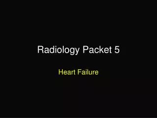
1 year MN Labrador Retriever“Baron” • Hx: Presented for evaluation of severe hip dysplasia. It is also noted that the patient is stiff in the elbows and stifles.
1 year MN Labrador Retriever“Baron” • RF • There is bilateral coxofemoral subluxation with degenerative joint changes. • The femoral heads are flattened and somewhat mushroom shaped. • The femoral necks are thickened and osteophyte formation is present. • The acetabulae are shallow and there is rounding of their cranial margins. • The lateral fabellae are large and irregularly shaped. • There is severe degenerative joint changes in the right elbow. • Osteophyte formation is present on the proximal radius, proximal ulna and distal humerus. • A very large osseous fragment is present in the cranial aspect of the joint. • There is prominent osteophyte formation on the medial coronoid process of the ulna seen on the oblique view. • The joint spaces are not well defined and there is sclerosis of the subchondral bone. • RD • Degenerative joint disease • Hip and elbow dysplasia
14-year old MN Shetland Sheepdog“Jet” • Hx: Presented for evaluation of hindlimb lameness. He has hyperflexion of his tarsi with palpable soft tissue thickening and crepitus of both joints.
14-year old MN Shetland Sheepdog“Jet” • RF • In the right tarsus there is subluxation at the level of the tarsometatarsal joint. • Focal punctate areas of bone lysis are seen in the adjacent subchondral bone surfaces at the tarsometatarsal joint. • There is marked thickening of the soft tissue surrounding the bone lesion, particularly on the lateral. • Remodeling of the distal end of the 2nd metatarsal bone is present. • Changes similar to those noted in the right tarsus are present in both carpi at the carpometacarpal joint. • There is irregular focal bone lysis in the subchondral bone with evidence of collapse of the joint spaces. • A focal area of irregular periosteal response is present on the right distomedial radius. • A smooth slightly irregular periosteal response is seen throughout the length of the 5th metacarpal bone. • Arthritic change is present at many of the metacarpophalangeal joints and at several interphalangeal joints. • Periosteal proliferation is present on the margins of the phalanges of the 5th digit bilaterally. • There is thickening of the soft tissues at the level of the carpi and surrounding the digits.
14-year old MN Shetland Sheepdog“Jet” • RD • Rheumatoid arthritis in the tarsometatarsal and carpometacarpal joints bilaterally
3-year old M Akita“Taiko” • Hx: Surgical repair of cranial cruciate ligament rupture 8 weeks ago. The sugical technique used was a fascia lata graft procedure. This is an intra-capsular technique. Taiko has now been lame for 3 weeks. The stifle is quite swollen and painful on palpation.
3-year old M Akita“Taiko” • RF • There is marked distension of the joint capsule as indicated by the decrease in size of the infrapatellar fat pad and caudal displacement of the gastrocnemius fascial plane. • Extra-capsular thickening of the soft tissue is present. • Osteophyte formation is present on the apex of the patella and the lateral aspect of the distal femur. The osteophytes are irregular and indistinctly seen indicating that this is an active process. • A metallic implant and bone cement from a prior total hip replacement are visible in the medullary cavity of the femur. • RD • Excessive soft tissue swelling with reactive bone changes associated with the patella and distal femur • R/O • Septic arthritis
13-year old mixed breed dog“Benji” • Hx: Presented with left forelimb lameness and shoulder pain.
13-year old mixed breed dog“Benji” • RF • Osteophyte formation of the periarticular margins of the glenoid cavity and the caudal aspect of the humeral head is present in both views. • There are multifocal bony opacities overlying the intertubercular groove of the humerus. • The subchondral bone of the humeral head and glenoid is sclerotic. • The contour of the caudal humeral head is mildly flattened. • RD • Severe degenerative joint disease of the shoulder • Mineralization of the biceps tendon, or sheath, or osteochondral fragments within the bicipital bursa
3-year old MN DLH“Diesel” • Hx: Left hindlimb lameness, decreased range of motion in the left tarsus and right carpus.
3-year old MN DLH“Diesel” • RF • There is significant subchondral lysis present within the talus, calcaneus and central tarsal bone. • There is arthritic change of the tibiotarsal, talocalcaneal, calcanoquartal and talocentral joints. • The proximal intertarsal joint space is collapsed. • There is mild thickening of the soft tissues in the region. • RD • Septic arthritis
3-year old MN Labrador Retriever“Buddy” • Hx: Chronic left cruciate ligament rupture with suspected meniscal damage.
3-year old MN Labrador Retriever“Buddy” • RF • There is moderate distension of the joint capsule as indicated by decrease in size of the infrapatellar fat pad and caudal displacement of the gastrocnemius fascial plane. • Mild degenerative joint disease is present. • Osteophyte formation is noted on the apex (distal end) of the patella, trochlear ridges of the femur and in the lateral and medial margins of the proximal tibia. • Faint areas of lucency in the proximal tibia seen in the CC view are evidence of subchondral lysis. • RD • Capsular distension with mild degenerative joint disease. • R/O • Cruciate ligament rupture.
1-month old Labrador Retriever • Hx: Presented with a swollen right tarsus, febrile.
1-month old Labrador Retriever • RF • Swelling of the right tarsus, severe. • Comparison of the right and left distal tibial epiphyseal bone shows some early destruction of the right. • Swelling also appears to be present around the right stifle. • RD • Septic arthritis of the right tarsus, probably the right stifle too • Next • Joint aspirate for culture and sensitivity