Hernia
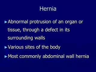
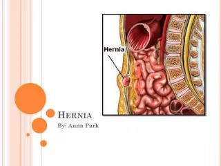
Hernia. Definition : A hernia is a protrusion of a viscus or a part of viscus through an abnormal opening in the walls of its containing cavity.

Hernia
E N D
Presentation Transcript
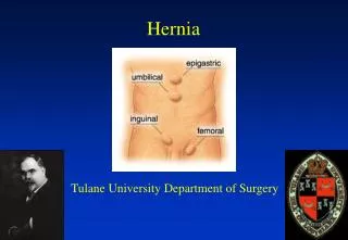
Definition : A hernia is a protrusion of a viscus or a part of viscus through an abnormal opening in the walls of its containing cavity. The external abdominal hernia is the most common form. The most frequent varieties are the inguinal, femoral and umbilical.
General features common to all hernias : Aetiology: Any condition that increase the intra – abdominal pressure such as a powerful muscular effort may produce a hernia. Whooping cough is a predisposing cause in childhood, whereas a chronic cough, straining on micturition or straining on defecation may precipitate a hernia in an adult. It should be remembered that appearance of hernia in an adult can be a sign of intra – abdominal malignancy. Obesity is an another factor, fat acts to separate muscle bundles and layers, weakens apponeurosis and leads to appearance of hernia ( para-umbilical, direct inguinal and hiatus hernia ). Hernia is more common in smokers which is due to acquired collagen deficiency increasing the risk of hernia
Compisition of a hernia: Hernia consists of three parts : sac, coverings and the contents. Sac : It is a diverticulum of peritoneum consisting of : 1 – Mouth. 2 – Neck. 3 – Body. 4 - Fundus. The neck is usually well defined but in some direct inguinal hernias and in many incisional hernias there is no actual neck. The diameter of the neck is important because strangulation of bowel is a likely complication when the neck is narrow as in femoral hernia and para-umbilical hernias. Covering : Derived from the layers of the abdominal wall through which the sac passes.
Contents : These can be : – Omentum = omentocele. – Intestine = interocele ( maily the small bowel but may be the large bowel ). – A portion of the circumference of the bowel = Richter’s hernia. – A portion of the bladder. – Ovary with or without the corresponding tube. – A Meckel’sdiverticulum = Littre’s hernia. – Fluid.
Classification : Irrespective of site, a hernia can be classified into 5 different types : 1 – Reducible. 2 – Irreducible. 3 – Obstructed. 4 – Strangulated. 5 – Inflammed. Reducible hernia : hernia either reduce itself when the patient lies down or can be reduced by the patient or the surgeon. Such a hernia ( reducible ) gives an expansible impulse on coughing. Irreducible hernia : in case the contents cannot be returned to the abdomen but there is no evidence of other complications. It is usually due to adhesions between the sac and the contents or overcrowding within the sac. Any degree of irreducibility predisposes to strangulation.
Obstructed hernia : It is irreducible hernia containing intestine that is obstructed from without or within but there is no interference to the blood supply to the bowel. The symptoms (colicky abdominal pain and tenderness over the hernia site) but less severe and onset is more gradual than in strangulated hernias. Usually there is no clear distinction clinically between obstruction and strangulation and the safe course is to assume that strangulation is imminent and treat accordingly.
Incarcerated hernia The term ‘incarceration’ is often used loosely as an alternative to obstruction or strangulation but is correctly employed only when it is considered that the lumen of that portion of the colon occupying a hernial sac is blocked with faeces. In this case, the scybalous contents of the bowel should be capable of being indented with the finger, like putty.
Strangulated hernia : A hernia becomes strangulated when the blood supply of its content is seriously impaired, rendering the contents ischaemic. Gangrene may occur as early as 5-6 hours after the onset of the first symptoms. Although inguinal hernia is more common than femoral hernia, a femoral hernia is more likely to strangulate because of the narrowness of the neck and its rigid surrounds. Pathology : The intestinal blood supply is impaired. Initially, only the venous return is impeded, the wall of the intestine becomes congested and bright red with the transudation of serous fluid into the sac. As congestion increases the wall of the intestine becomes purple in color. The intestinal pressure increases, distending the intestinal lop and impairing venous return further. As venous stasis increases, the arterial supply becomes more and more impaired. Blood is extravasataed under the serosa and is effused into the lumen.
The fluid in the sac becomes blood stained and shining serosa dull because of a fibrinous, sticky exudate. At this stage the walls of the intestine have lost their tone and become friable. Bacterial transudation occurs secondary to the lowered intestinal viability and the sac fluid becomes infected. Gangrened appears at the rings of constriction, which becomes deeply indented and grey in color. The gangrene then then develops in the ant mesenteric border, the color varying from black to green depending on the decomposition of the blood in the subserosa. The mesentery involved by the strangulation also becomes gangerenous. If the strangulation is unrelieved, perforation of the wall of the intestine occurs, either at the convexity of the loop or at the seat of constriction. Peritonitis spreads from the sac to the peritoneal cavity.
Clinical features : Sudden pain at first situated over the hernia, is followed by generalized abdominal pain, colicky in character and often located mainly at the umbilicus. Nausea and subsequently vomiting. The patient may complain of an increase in hernia size. On examination the hernia is tense, extremely tender and irreducible and no expansile cough impulse. Unless the strangulation is relieved by operation, the spasms of pain continue until peristaltic contractions cease with the onset of ischemia, when paralytic ileus ( often the result of peritonitis ) and septicaemia develops. Spontaneous cessation of pain must be viewed with caution, as this may be a sign of perforation
Strangulated hernia ■ Present with local then general abdominal pain and vomiting ■ A normal hernia can strangulate at any time ■ Most common in hernias with narrow necks such as femoral hernias ■ Require urgent surgery
Imflammed hernia : inflammation can occur from inflammation of the contents of the sac as eg. acute appendicitis or salpingitis. Inflammation can happen from external causes eg. Trophic ulcers that developes in the depending areas of large umbilical or incisional hernias. The hernia is usually tender but not tense and the overlying skin red and oedematous. Treatment is based on treatment of the inderlying cause.
Inguinal Hernia Surgical anatomy : The superficial inguinal ring is a triangular aperature in the aponeurosis of the external oblique muscle lies 1.25 cm above the pubic tubercle. The deep inguinal ring is a U shaped condesation of the transervalis fascia and it lies 1.25 cm above the inguinal ligament
An indirect hernia travels down the canal on the outer side of the spermatic cord. A direct hernia comes out directly forwards through the posterior wall of the inguinal canal. Whereas the neck of the indirect hernia is lateral to the inferior epigastric artery, the neck of the direct hernia is medial to the artery. An inguinal hernia can be differentiated from femoral hernia by the ascertaining the relation of the neck of the sac to the medial end of the inguinal ligament and the pubic tubercle. In the case of an inguinal hernia, the neck is above and medial, whereas that of a femoral hernia is below and lateral
Indirect inguinal hernia It is the most common type of hernia. It is more common in the young while the direct type is more common in the old. In the first decade of life, inguinal hernia is more common on the right side in the male. This is associated with the later descent of the right testis and a higher incidence of failure of closure of the process vaginalis. Clinical features : We start examining the patient in standing position with asking the patient to cough and feel the cough impulse. Then examiner should now : * Is the hernia is right, left or bilateral. * Is it inguinal or femoral. * Is it direct or indirect. * Is it reducible or not. * Is it complete or not. * What are the contents
The patient complains of pain in the groin or pain referred to the testicles when performing heavy wok or taking strenuous exercise. In large hernias there is a sensation of weight and dragging on the mesentery which may produce epigastric pain. Differential diagnosis in the male : 1 – Vaginal hydrocele. 2 – Encysted hydrocele of the cord. 3 – Spermatocele. 4 – Femoral hernia. 4 – Incompletely descended testis. 5 – Lipoma of the cord. Differential diagnosis in the female : 1 – Hydrocele of the canal of Nuck. 2 – Femoral hernia.
Note that examination using finger and thumb across the neck of the scrotum will help to distinguish between a swelling of inguinal origin and one that is entirely intrascrotal.
Treatment : Operation is the treatment of choice. ( Herniotomy+ Herniorrhaphy ) * Herniotomy : means dissecting out and opening the hernial sac, reducing any content then transfixing the neck of the sac and removing the remainder. By itself it is sufficient for the treatment of hernia in children and young adults. * Herniorrhaphy : consist of : 1 – repair of the stretches internal ring and transversalis fascia. 2 – further reinforcement of the posterior wall of the inguinal canal.
* A truss may be used when operation is contraindicated or refused. In this condition if the truss to be used the hernia should be reducible. Its use should be mainly historical, as there are very few contraindications to surgery with today’s variety of anaesthetic techniques.
Direct inguinal hernia It is always acquired. The passes through a weakness or a defect of the transversalis fascia in the posterior wall of the inguinal canal. Predisposing factors are smoking and occupations that involves straining or heavy lifting. Damage to the ilioinguinal nerve (previous appendicectomy) is another cause, because of the resulting weakness of the conjoined tendon. Direct hernia do not often attain a large size or reach to the scrotum. In contrast to the indirect hernia, the direct lies behind the spermatic cord. As the neck is wider than that of indirect inguinal hernia, direct inguinal hernia do not often strangulate. Treatment : the principles of repair of direct hernia are the same as those of an indirect hernia, with the exception that the hernia sac can usually be simply inverted after it has been dissected free and the transversalis fascia reconstructed in front of it. This repair can be done also by mesh which is also true for indirect hernia repair.