Download
1 / 33
330 likes | 378 Views
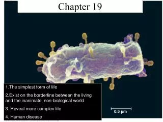
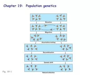
Fig. 19-1. 0.5 µm. RESULTS. Fig. 19-2. Extracted sap from tobacco plant with tobacco mosaic disease. Passed sap through a porcelain filter known to trap bacteria. Rubbed filtered sap on healthy tobacco plants. 3. 1. 2. Healthy plants became infected. 4. RNA. Membranous

E N D
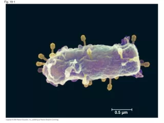
Fig. 19-1 0.5 µm
RESULTS Fig. 19-2 Extracted sap from tobacco plant with tobacco mosaic disease Passed sap through a porcelain filter known to trap bacteria Rubbed filtered sap on healthy tobacco plants 3 1 2 Healthy plants became infected 4
RNA Membranous envelope Head DNA RNA DNA Capsomere Fig. 19-3 Capsid Tail sheath Tail fiber Capsomere of capsid Glycoproteins Glycoprotein 18 250 nm 70–90 nm (diameter) 80 225 nm 80–200 nm (diameter) 20 nm 50 nm 50 nm 50 nm (a) Tobacco mosaic virus (b) Adenoviruses (d) Bacteriophage T4 (c) Influenza viruses
RNA Fig. 19-3a Capsomere of capsid 18 250 nm 20 nm (a) Tobacco mosaic virus
DNA Capsomere Fig. 19-3b Glycoprotein 70–90 nm (diameter) 50 nm (b) Adenoviruses
Membranous envelope RNA Capsid Fig. 19-3c Glycoproteins 80–200 nm (diameter) 50 nm (c) Influenza viruses
Head DNA Fig. 19-3d Tail sheath Tail fiber 80 225 nm 50 nm (d) Bacteriophage T4
VIRUS Entry and uncoating 1 DNA Capsid Transcription and manufacture of capsid proteins 3 Fig. 19-4 Replication 2 HOST CELL Viral DNA mRNA Capsid proteins Viral DNA Self-assembly of new virus particles and their exit from the cell 4
Attachment 1 Fig. 19-5-1
Attachment 1 2 Entry of phage DNA and degradation of host DNA Fig. 19-5-2
Attachment 1 2 Entry of phage DNA and degradation of host DNA Fig. 19-5-3 3 Synthesis of viral genomes and proteins
Attachment 1 2 Entry of phage DNA and degradation of host DNA Fig. 19-5-4 Phage assembly 4 Assembly 3 Synthesis of viral genomes and proteins Head Tail Tail fibers
Attachment 1 2 Entry of phage DNA and degradation of host DNA Fig. 19-5-5 5 Release Phage assembly 4 Assembly 3 Synthesis of viral genomes and proteins Head Tail Tail fibers
Daughter cell with prophage Fig. 19-6 Phage DNA The phage injects its DNA. Cell divisions produce population of bacteria infected with the prophage. Phage DNA circularizes. Phage Bacterial chromosome Occasionally, a prophage exits the bacterial chromosome, initiating a lytic cycle. Lytic cycle Lysogenic cycle The bacterium reproduces, copying the prophage and transmitting it to daughter cells. The cell lyses, releasing phages. Lytic cycle is induced Lysogenic cycle is entered or Prophage Phage DNA integrates into the bacterial chromosome, becoming a prophage. New phage DNA and proteins are synthesized and assembled into phages.
Capsid and viral genome enter the cell Capsid RNA Fig. 19-7 HOST CELL Envelope (with glycoproteins) Viral genome (RNA) Template mRNA Capsid proteins ER Copy of genome (RNA) Glyco- proteins New virus
Viral envelope Glycoprotein Capsid RNA (two identical strands) Reverse transcriptase HIV Fig. 19-8 Membrane of white blood cell HIV HOST CELL Reverse transcriptase Viral RNA RNA-DNA hybrid 0.25 µm DNA HIV entering a cell NUCLEUS Provirus Chromosomal DNA RNA genome for the next viral generation mRNA New virus New HIV leaving a cell
Viral envelope Glycoprotein Capsid RNA (two identical strands) Fig. 19-8a HOST CELL Reverse transcriptase HIV Reverse transcriptase Viral RNA RNA-DNA hybrid DNA NUCLEUS Provirus Chromosomal DNA RNA genome for the next viral generation mRNA New virus
Membrane of white blood cell HIV Fig. 19-8b 0.25 µm HIV entering a cell New HIV leaving a cell
Fig. 19-9 (a) The 1918 flu pandemic 0.5 µm (b) Influenza A H5N1 virus (c) Vaccinating ducks
Fig. 19-9a (a) The 1918 flu pandemic
Fig. 19-9b 0.5 µm (b) Influenza A H5N1 virus
Fig. 19-9c (c) Vaccinating ducks
Fig. 19-11 Original prion Prion Aggregates of prions New prion Normal protein
The phage attaches to a host cell and injects its DNA Phage DNA Fig. 19-UN1 Bacterial chromosome Prophage Lytic cycle Lysogenic cycle • Virulent or temperate phage • Destruction of host DNA • Production of new phages • Lysis of host cell causes release • of progeny phages • Temperate phage only • Genome integrates into bacterial • chromosome as prophage, which • (1) is replicated and passed on to • daughter cells and • (2) can be induced to leave the • chromosome and initiate a lytic cycle
Fig. 19-UN2 B A Number of viruses Number of bacteria Time Time