Glycosaminoglycans in Cell Biology
340 likes | 440 Views
Learn about the structure and functions of glycosaminoglycans, heparan sulfate, proteoglycan complexes, collagen fibers, and the extracellular matrix. Explore the physical properties, biosynthesis, and role in cell processes.

Glycosaminoglycans in Cell Biology
E N D
Presentation Transcript
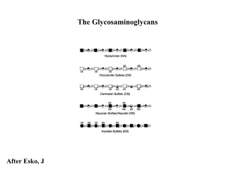
The Glycosaminoglycans After Esko, J
Heparan sulphate structure Proteoglycan complex Collagen Fiber Proteoglycan molecules form complexes by noncovalently attaching to long polysaccharide molecules Polysaccharide molecule Proteoglycan molecule Collagen fibers are embedded in a web of proteoglycan complexes Plasma membrane Integrin Integrins are membrane proteins that are bound to the ECM on one side and to microfilaments on the cytoskeleton on the other. This linkage can transmit stimuli between the cell’s external environment and its interior. Fibronectins attach the ECM to the plasma membrane of the cell Components of the extracellular matrix Microfilaments of cytoskeleton • Anionic polysaccharide chains • Fibrous proteins
b4 b3 b 4 b 3 b 4 b 3 b 4 b 3 b 4 b 3 Hyaluronan (HA) n≥1000 GlcNAc GlcA GlcNAc GlcA GlcNAc GlcA GlcNAc GlcA GlcNAc GlcA • Abundant in skeletal tissues, synovial fluid, skin, and vitreous • Ovulation/ fertilization • Macrophage stimulation • Angiogenesis • Cell migration • Morphogenesis and differentiation After Esko, J
Karl Meyer, Columbia University Simoni, R. D. et al. J. Biol. Chem. 2002;277:e27
Physical Properties • Gels of high viscosity, and a great lubricant since at high shear its viscosity drops, but remains resilient • Interglycosidic H-bonding restricts rotations across glycosidic bonds • Promotes rapid recovery after mechanical perturbations • Hydrated matrices rich in hyaluronan expand the extracellular space, facilitating cell migration. • There is both a polar and a hydrophobic face for interaction with other macromolecules After Hascall and Laurent
HA synthase(s) located in plasma membrane • Copolymerization of UDP-GlcNAc and UDP-GlcA occurs independently of a core protein • HA can contain 250-25,000 disaccharides (105- 4x 10⁷ Da, ~10 µm, the length of an erythrocyte) • Half-life rate ranges from 2 weeks in synovial fluid to 5 minutes in the bloodstream After Weigel, P
Diagram of a putative metabolic scheme for hyaluronan degradation. From Stern, R.
Hyaluronan Binding Proteins After Esko, J
Aggrecan Hyaluronic Acid • High charge density creates osmotic pressure that draws water into the tissue (sponge) • Absorbs high compressive loads, yet resilient Cartilage - Proteoglycan Aggregates • Aggrecan: Large chondroitin sulfate proteoglycan present in cartilage and other connective tissues • Core protein ~400 kDa • ~100 chondroitin sulfate chains of ~20 kDa • Forms aggregates with hyaluronic acid (HA) After Esko, J
Albert Dorfman, University of Chicago Kresge, N. et al. J. Biol. Chem. 2005;280:e28
b4 b3 b 4 b 3 b 4 b 3 b 4 b 3 b 4 b 3 Chondroitin Sulfate IdoA 6S 6S 6S 4S 4S 4S GalNAc GlcA Non-sulfated chondroitin is rare in vertebrates, but multiple types of sulfated chondroitins are known (A, B, C, D, etc) Multiple sulfotransferases decorate the chain An epimerase can flip the stereochemistry of D-GlcA to L-IdoA (Dermatan Sulfate) The chains are easily characterized using bacterial chondroitinases which degrade the chain to disaccharides After Esko, J
6 6 S S 6 S N S N S N S 2 S N S 3 S Heparin/Heparan Sulfate IdoA GlcNAc GlcA Gal Gal Xyl • Characterization of heparan sulfate is based on different criteria • - GlcNAc vs GlcNS • - 3-O-Sulfo and 6-O-sulfo groups • -IdoA vs GlcA • Heparinases degrade chain into disaccharide units • Nitrous acid degrades chains at GlcNS • Disaccharides characterized by HPLC or mass spectrometry After Esko, J
Copolymerase Complex EXT1/EXT2 GlcNAc/S 6-O-sulfotransferases (6OST) (3+ isozymes) EXTL2/3 Epimerase IdoA GlcNAc 6 S 6 S 6 S N S N S N S N 2 S S GlcA Gal Gal Xyl 3 S GlcNAc N-deacetylase N-sulfotransferases (NDST) (4 isozymes) Uronic acid 2-O-sulfotransferase GlcNH2/S 3-O-sulfotransferases (3OST) (6 isozymes) Biosynthesis of a Heparin/Heparan Sulfate Chain After Esko, J
6S 6S 6S NS 2S NS 2S NS 6S 6S 6S NS NS 2S NS 3S NS NS NS 4S GlcA 4S 4S 4S 4S GlcNAc IdoA Chondroitin sulfate Heparan Sulfate Proteoglycans After Esko, J
The Heparin Anti-thrombin III binding motif Lindahl, U
Heparin’s function in the mast cell appears to have nothing to do with disturbed blood coagulation. Heparin is discharged from the mast cell only in special emergency situations, such as anaphylactic shock or other inflammatory reactions. • Marine mussels have no blood in the conventional sense to anticoagulate, yet the polysaccharide in the "mast cells" turns out to contain the specific antithrombin-binding pentasaccharide sequence. It also has very high anticoagulant activity. It seems reasonable to assume that the clams contain a protease inhibitor related to antithrombin (belonging to the serpin family), but for a functional reason quite unrelated to blood coagulation.
S h e d d i n g H S c h a i n P l a s m a m e m b r a n e E n d o c y t o s i s S t e p 1 p r o t e a s e e n d o g l y c o s i d a s e G o l g i H S o l i g o s a c c h a r i d e S t e p 2 ( ~ 1 0 k D a ) e n d o g l y c o s i d a s e H S o l i g o s a c c h a r i d e ( ~ 5 k D a ) S t e p 3 e x o g l y c o s i d a s e s u l f a t a s e Proteoglycan Turnover • Shedding by exoproteolytic activity, MMP-7 for one • Endosulfatases remove sulfate groups on proteoglycans at cell surface: remodeling • Heparanase (endohexosaminidase) clips at certain sites in the chain. Outside cells, it plays a role in cell invasion processes • Inside cells it’s the first step towards complete degradation in lysosomes by exoglycosidases and sulfatases Mucopolysaccharidoses After Esko, J
Mucopolysaccharidoses - Lysosomal Storage diseases • I H: Hurler's a-L-iduronidase corneal clouding; dwarfism; mental retardation; early mortality • MPS I S: Scheie's a -L-iduronidase corneal clouding; aortic valve disease; joint stiffening; normal intelligence and life span • MPS I H/S: Huler/Scheie a -L-iduronidase similar to both I-H and I-S • MPS II: Hunter's L-iduronate-2-sulfatase mild and severe forms, X-linked, also a possible autosomal form, facial and physical deformities; mental retardation • MPS III A: Sanfilippo(A) Heparan N-sulfatase skin, brain, lungs, heart and skeletal muscle are affected in all 4 types of MPS-III • MPS III B: Sanfilippo(B)N-acetyl-a -D-glucosaminidase congestive heart failure; progressive mental retardation • MPS III C: Sanfilippo(C)N-acetylCoA: a-glucosamine- N-acetyltransferase coarse facial features; organomegaly • MPS III D: Sanfilippo(D)N-acetyl-a -glucosamine-6-sulfatase moderate physical deformities; progressive mental retardation • MPS IV A: Morquio's(A) N-acetylgalactosamine- 6-sulfatase corneal clouding, thin enamel, aortic valve disease, skeletal abnormalities • MPS IV B: Morquio's(B) ß -galactosidasemild skeletal abnormalities, normal enamel, hypoplastic odontoid, corneal clouding • MPS VI Maroteaux-Lamy N-acetylgalactosamine- 4-sulfatase 3 distinct forms from mild to severe, aortic valve disease, normal intellect, corneal clouding, coarse facial features • MPS VII Sly ß –glucuronidase hepatosplenomegaly, dystosis multiplex Michael W. KING, IU School of Medicine
Keratan sulfate I (corneal) and II (skeletal) Figure 11.4. Keratan sulfates consist of a sulfated polylactosamine linked either to Asn or Ser/Thr residues. The actual order of the various sulfated and nonsulfated disaccharides occurs somewhat randomly along the chain. Not shown are sialic acid and fucose residues that may be present at the termini of the chains.
GAG Summary • Proteoglycans contain glycosaminoglycans: chondroitin sulfate, dermatan sulfate, or heparan sulfate • Heparan sulfate, Chondroitin, and dermatan sulfate proteoglycans are found in the matrix and play structural roles in cartilage, bone and soft tissues and are found at the cell surface and play roles in cell adhesion and signaling during development • Proteoglycans in the extracellular matrix can also act as a reservoir of growth factors, protect growth factors from degradation, and facilitate the formation of gradients • Human diseases in proteoglycan assembly are rare • Degradation of these compounds is also important (MPS) After Esko, J
Functional roles of GAGs • GAGs have long been viewed as static, primarily structural molecules. Evidence from functional studies show that GAGs and the proteoglycans are highly dynamic, with rapid turnover rates. This provides them with essential roles in development, and differentiation, pathogenicity, and various pathologies.
Secreted PGs • Aggrecan CS/KS brain, cartilage • Versican CS brain, mesenchyme • Neurocan CS brain • Brevican*CS brain • Decorin CS/DS brain, connective tissue • Biglycan CS/DS brain, connective tissue • Testican CS/HS brain, testis • Perlecan HS brain, basement membrane • Dystroglycan HS brain, muscle • Agrin HS brain, muscle • Claustrin KS brain Glycoword
Transmembrane PGs • NG2-PG CS brain, cartilage • RPTP zeta/beta (phosphacan) CS/KS brain, cartilage • Neuroglycan C CS brain • Appican CS brain, connective tissue • N-syndecan HS brain • SV2 KS brain • GPI-anchored PGs • Glypican HS brain, muscle • K-glypican HS brain, kidney • Cerebroglycan HS brain Glycoword
Bamacan: Chondroitin Sulfate Proteoglycan Perlecan: Heparan Sulfate Proteoglycan Laminin Entactin/Nidogen Collagen Type IV Extracellular Matrices • Epithelial cells produce basement membranes composed of proteoglycans, reticular collagens and glycoproteins After Esko, J
HSPGs function as co-receptors for growth factors and their receptor tyrosine kinases, which are present either on the same cell (a) or on adjacent cells (b). They transport chemokines across cells (c) and present them at the cell surface (d). Proteolytic processing leads to the shedding of syndecans and glypicans from the cell surface (e), and heparanase cleaves the HS chains (f), liberating bound ligands (such as growth factors). Cell-surface HSPGs are actively taken up by endocytosis (g) and can recycle back to the surface or be degraded in lysosomes (h). HSPGs also facilitate cell adhesion to the extracellular matrix (i) and form bridges to the cytoskeleton (j). Secreted HSPGs are involved in the formation of organized extracellular matrices that form physiological barriers (k) and sequester growth factors and morphogens for later release (l). Serglycin carrying highly sulphated heparin chains is packaged into secretory granules of haematopoetic cells (m).
GAG-Chemokine interactions • Chemokines are small secreted proteins that are critically involved in many biological processes, including routine immune surveillance, inflammation and development. • Specific chemokine receptors provide the portals by which the HIV virus gets into cells, while others have been implicated in inflammatory diseases associated with inappropriate cell migration (rheumatoid arthritis, multiple sclerosis, atherosclerosis, and most recently, tumor metastasis). • HSPGs are involved in delivering, sequestering, and presenting chemokines. Potential roles of CSPGs as well, and variations in type of PG and degree of sulphation may play be critical factors in sequestration and presentation.
Figure 2. Schematic representation of P. falciparum VAR2CSA. Of the six DBL domains, DBL2-x, DBL3-x and DBL6-e are confirmed to bind to CSA. DBL6 ε - x DBL2x DBL2 - x DBL3 DBL3x - x DBL4ε DBL4 - ? DBL5ε DBL5 - ? DBL6 - ? DBL1x DBL1 Figure 1 Placental malaria • The malaria parasite Plasmodium falciparum expresses adhesive proteins on the surface of infected red blood cells that enable them to bind to host receptors on the endothelium and within the placenta. Encoded by the highly variant var multi-gene family, only one member has been demonstrated to mediate binding within the placenta. This placenta-specific adhesive protein, VAR2CSA, binds to a form of chondroitin sulfate A (CSA) unique to the placenta.
Role of Heparan in Slit-Robo interactions • Slits are large secreted and multidomain leucine-rich repeat (LRR)-containing glycoproteins with multiple roles in cell signaling and adhesion. • More recently, Slits have been implicated in direct cell migration, axonal guidance, and sensory axon elongation in vertebrate models, heart morphogenesis, angiogenesis, and tumor metastasis. • Slits are ligands for the Robo (Roundabout) receptors, which belong to the Ig superfamily of transmembrane signaling molecules. • The Slit-Robo interaction is mediated by the second LRR domain of Slit and the two N-terminal Ig domains of Robo, but the molecular details of this interaction and how it induces signaling remain unclear.
Composite heparin binding site in the Slit-Robo complex Fukuhara, N. et al. J. Biol. Chem. 2008;283:16226-16234
Role of CD44-HA interactions in leukocyte rolling and metastasis Primary adhesion or weak, intermittent adhesion under flow conditions apparently functions as a ‘braking system’ for flowing leukocytes and tumor cells in the circulation so that they can be trapped effectively to vascular surfaces during the course of lymphocyte trafficking into tissues [1] or of hematogenous metastasis of tumor cells [29]. This phenomenon has been shown to occur in the interactions between selectins and their carbohydrate ligands [1] and between CD44 and HA [2]. Toshiyuki Muraia, Nagako Sougawaa, Hiroto Kawashimaa, Kazuhito Yamaguchib
Roles of GAGs in neural development and disease • “Remarkably, the developmental changes in lectican expression correlate with changes in the biophysical properties of the ECM that affect neural plasticity. During early development, these CSPGs are associated but not densely aggregated to the HA scaffold, and compose an ECM with large, hydrated spaces that enables neural cell motility and axonal navigation. Conversely, the adult matrix contains dense, insoluble aggregates of HA and bound CSPGs that are deposited around neurons and glia, which provide cellular support but limit cellular motility and plasticity. It has been suggested that this change is caused by the shift in CSPG expression from a group of early predominant CSPGs, including neurocan, V1 versican and other non-lectican CSPGs [e.g. receptor protein tyrosine phosphatase (RPTP)-β/ζ/phosphacan and neuroglycan-C], to adult molecules (aggrecan, V2 versican and Brevican) [18]. The nature of the matrix would change owing to differences in amount, localization and molecular interactions of the juvenile CSPGs, which would enable cell and/or process movement, and those of the adult CSPGs, which would aggregate around cells and neurites and limit their ability for spatial relocation. Similarly, changes in the structures and expression patterns of the CSPGs in neuropathologies might correspond to changes in the properties and functions of the neural matrix in the diseased CNS. “ • Mariano S. Viapianoa and Russell T. Matthews • Formation of perineural nets • Alzheimers- formation of amyloid plaques • Gliomas