Download
1 / 14
140 likes | 267 Views
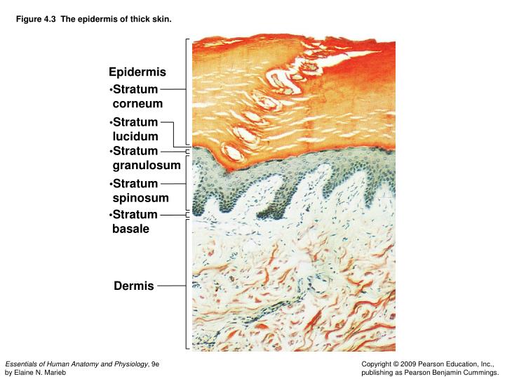
Figure 4.3 The epidermis of thick skin. Epidermis. Stratum corneum. Stratum lucidum. Stratum granulosum. Stratum spinosum. Stratum basale. Dermis. Figure 4.3 The epidermis of thick skin.

E N D
Figure 4.3 The epidermis of thick skin. Epidermis • Stratum • corneum • Stratum • lucidum • Stratum • granulosum • Stratum • spinosum • Stratum • basale Dermis
Figure 4.7a Structure of a hair and hair follicle. (a) Longitudinal section of a hair within its follicle. Hair shaft Arrector pili Sebaceous gland Hair root Hair bulb in follicle (a)
Figure 4.7a Structure of a hair and hair follicle. (a) Longitudinal section of a hair within its follicle. (a)
Figure 4.7b Structure of a hair and hair follicle. (b) Hair. Cuticle Cortex Medulla (b) Hair
Figure 4.7b Structure of a hair and hair follicle. (b) Hair. (b) Hair
Figure 4.7c Structure of a hair and hair follicle. (c) Enlarged longitudinal view of the expanded hair bulb in the follicle showing the matrix, the region of actively dividing epithelial cells that produces the hair. Dermal sheath Hair follicle Epidermal sheath Matrix (growth zone) in hair bulb Melanocyte Connective tissue papilla containing blood vessels (c)
Figure 4.7c Structure of a hair and hair follicle. (c) Enlarged longitudinal view of the expanded hair bulb in the follicle showing the matrix, the region of actively dividing epithelial cells that produces the hair. (c)
Figure 4.9a Structure of a nail. (a) Surface view. Body of nail Lateral nail fold Lunula Cuticle (a) Surface view
Figure 4.9a Structure of a nail. (a) Surface view. Surface view
Figure 4.9b Structure of a nail. (b) Longitudinal section of the distal part of a finger. Nail matrix Root of nail Proximal nail fold Nail bed Free edge of nail Cuticle Body of nail Bone of fingertip Stratum basale (b) Longitudinal section of the distal part of a finger
Figure 4.9b Structure of a nail. (b) Longitudinal section of the distal part of a finger. (b) Longitudinal section of the distal part of a finger
Appendages of the Skin Figure 4.6b
Appendages of the Skin Figure 4.6a