Download
1 / 109
1.15k likes | 1.39k Views
Organization of the Cell. Chapter 4. Learning Objective 1. What is cell theory ? How does cell theory relate to the evolution of life?. Cell Theory. Cells are basic units of organization and function in all living organisms All cells come from other cells

E N D
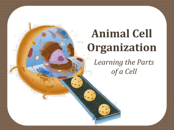
Organization of the Cell Chapter 4
Learning Objective 1 • What is cell theory? • How does cell theory relate to the evolution of life?
Cell Theory • Cells are basic units of organization and function in all living organisms • All cells come from other cells All living cells have evolved from a common ancestor
Learning Objective 2 • What is the relationship between cell organization and homeostasis?
Homeostasis • Cells have many organelles, internal structures that carry out specific functions, that help maintain homeostasis
KEY CONCEPTS • Cell organization and size are critical in maintaining homeostasis
Plasma Membrane • Plasma membrane • surrounds the cell • separates cell from external environment • maintains internal conditions • allows the cell to exchange materials with outer environment
KEY CONCEPTS • Eukaryotic cells are divided into compartments by internal membranes • Membranes provide separate, small areas for specialized activities
Learning Objective 3 • What is the relationship between cell size and homeostasis?
Mitochondrion Red blood cells Human egg Chloroplast Typical bacteria Chicken egg Protein Virus Amino acids Nucleus Atom Smallest bacteria Epithelial cell Adult human Ribosomes Frog egg Some nerve cells 0.1 nm 1 nm 10 nm 100 nm 1 μm 10 μm 100 μm 1 mm 10 mm 100 mm 1 m 10 m Electron microscope Light microscope Human eye Measurements 1 meter = 1000 millimeters (mm) 1 millimeter = 1000 micrometers (μm) 1 micrometer = 1000 nanometers (nm) Fig. 4-1, p. 75
Surface to Volume Ratio • SVR • ratio of plasma membrane (surface area) to cell’s volume • regulates passage of materials into and out of the cell • Critical factor in determining cell size
1 mm 2 mm 2 mm 1 mm Surface area = height width number of sides number of cubes 48 Surface Area (mm2) 24 (2 2 6 1) (1 1 6 8) Volume = height width length number of cubes 8 Volume (mm3) 8 (2 2 2 1) (1 1 1 8) Surface Area/ Volume Ratio Surface area/ volume 3 6 (48 :8) (24 :8) Fig. 4-2, p. 76
Learning Objective 4 • What methods do biologists use to study cells? • How are microscopy and cell fractionation used?
Microscopes • Light microscopes • Electron microscopes • superior resolving power
Light microscope Light beam Ocular lens Objective lens Specimen Condenser lens Light source (a) A phase contrast light microscope can be used to view stained or living cells, but at relatively low resolution. 100 μm Fig. 4-4a, p. 79
Transmission electron microscope Electron gun Electron beam First condenser lens (electromagnet) Specimen Projector lens (electromagnetic) Film or screen (b) The transmission electron microscope (TEM) produces a high-resolution image that can be greatly magnified. A small part of a thin slice through the Paramecium is shown. 1 μm Fig. 4-4b, p. 79
Scanning electron microscope Electron gun Electron beam Second condenser lens First condenser lens (electromagnet) Scanning coil Final (objective) lens Cathode ray tube synchronized with scanning coil Secondary electrons Specimen Electron detector (c) The scanning electron microscope (SEM) provides a clear view of surface features. 100 μm Fig. 4-4c, p. 79
Cell Fractionation • Cell fractionation • purifies organelles • to study function of cell structures
Centrifuge rotor Centrifugal force Centrifugal force Hinged bucket containing tube (a) Centrifugation. Due to centrifugal force, large or very dense particles move toward the bottom of a tube and form a pellet. Fig. 4-5a, p. 80
Layered microsomal suspension Centrifuge supernatant 20,000 x G Low sucrose concentration Centrifuge supernatant 100,000 x G Plasma membrane Centrifuge 600 x G Density gradient centrifugation Resuspend pellet layer on top of sucrose gradient 10 minutes 30 minutes 90 minutes 100,000 x G Sucrose density gradient High Golgi sucrose concentration Nuclei in pellet Mitochondria, chloroplasts in pellet Disrupt cells in buffered solution Microsomal pellet (contains ER, Golgi, plasma membrane) ER (b) Differential centrifugation. Cell structures can be separated into various fractions by spinning the suspension at increasing revolutions per minute. Membranes and organelles from the re-suspended pellets can then be further purified by density gradient centrifugation (shown as last step). G is the force of gravity. ER is the endoplasmic reticulum. Fig. 4-5b, p. 80
Layered microsomal suspension Low sucrose concentration Centrifuge supernatant 20,000 x G Centrifuge supernatant 100,000 x G Plasma membrane Density gradient centrifugation Centrifuge 600 x G Resuspend pellet layer on top of sucrose gradient Sucrose density gradient 10 minutes 90 minutes 30 minutes 100,000 x G Disrupt cells in buffered solution Golgi Nuclei in pellet Mitochondria, chloroplasts in pellet Microsomal pellet (contains ER, Golgi, plasma membrane) High sucrose concentration ER Stepped Art Fig. 4-5b, p. 80
Learning Objective 5 • How do the general characteristics of prokaryotic and eukaryotic cells differ? • How are plant and animal cells different?
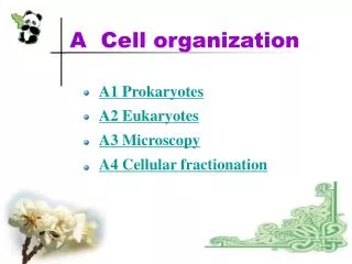
Prokaryotes • Prokaryotic cells • No internal membrane organization • nuclear area (not nucleus) • cell wall • ribosomes • flagella
Pili Storage granule Flagellum Ribosome Cell wall DNA Plasma membrane Nuclear area Capsule 0.5 μm Fig. 4-6, p. 81
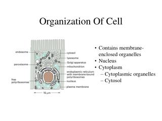
Eukaryotes • Eukaryotic cells • membrane-enclosed nucleus • cytoplasm contains organelles • cytosol (fluid component)
Chromatin Membranous sacs of Golgi Nuclear envelope Nucleolus Nuclear pores Golgi complex Nucleus Plasma membrane Lysosome Nuclear envelope Cristae Ribosomes Rough ER Rough and smooth endoplastic reticulum (ER) Centrioles Mitochondrion Smooth ER Fig. 4-8, p. 83
Plant Cells • Plant cells • rigid cell walls • plastids • large vacuoles • no centrioles
Mitochondrion Cristae Membranous sacs Golgi complex Cell wall Plasma membrane Vacuole Chloroplast Nucleus Granum Smooth ER Nuclear envelope Stroma Nucleolus Nuclear pores Rough ER Chromatin Ribosomes Rough and smooth endoplasmic reticulum (ER) Fig. 4-7, p. 82
Learning Objective 6 • What are the three functions of cell membranes?
Cell Membranes • Divide cell into compartments • Vesicles transport materials between compartments • Important in energy storage and conversion • Endomembrane system
Learning Objective 7 • What are the structures and functions of the nucleus?
The Nucleus • Control center of cell • genetic information coded in DNA • Nuclear envelope • double membrane • Nuclear pores • communicate with cytoplasm
Nuclear Structures • Chromatin • DNA and protein • Chromosomes • DNA condensed for cell division • Nucleolus • ribosomal RNA synthesis • ribosome assembly
Rough ER Chromatin Nuclear pores Nucleolus (b) 0.25 μm Nuclear envelope Nuclear pore ER continuous with outer membrane of nuclear envelope Nucleoplasm Outer nuclear envelope Nuclear pore 2 μm (a) Nuclear pore proteins Inner nuclear envelope (c) Fig. 4-11, p. 88
KEY CONCEPTS • Eukaryotic cells have nuclei containing genetic information coded in DNA
Learning Objective 8 • What are the structural and functional differences between smooth ER and rough ER?
Endoplasmic Reticulum (ER) • Network of folded membranes • in cytosol • Smooth ER • lipid synthesis • calcium ion storage • detoxifying enzymes • Rough ER • ribosomes on outer surface • assembles proteins
ER lumen Mitochondrion Ribosomes Rough ER 1 μm Smooth ER Fig. 4-12, p. 90
Learning Objective 9 • Trace the path of protein synthesis: • synthesis in the rough ER • processing, modification, and sorting by the Golgi complex • transportation to specific destinations
The Golgi Complex • Processes proteins synthesized by ER • Manufactures lysosomes • Cisternae • stacks of flattened membranous sacs
Transport Vesicles • Formed by membrane budding • Move glycoproteins • from ER to cis face of Golgi complex • Carry modified proteins from trans face to specific destination