Download
1 / 72
720 likes | 746 Views
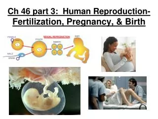
Learn about the intricate process of fertilization and early embryo development, outlining key steps from acrosome reaction to blastocyst implantation, and the vital role of the placenta in maternal-fetal exchange during pregnancy.

E N D
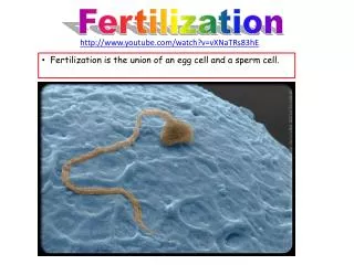
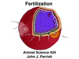
IB Assessment Statement • Describe the process of fertilization, including the acrosome reaction, penetration of the egg membrane by a sperm and the cortical reaction.
Fertilization • Fertilisation occurs in the oviducts (fallopian tubes) • pH of vagina is acidic and pH of semen is basic. Thus they neutralize each other.
Fertilization • Contraction of the uterus and oviducts help move the sperm into the oviducts. • One or more sperm will reach the oocyte in the oviduct.
Fertilisation • The oocyte is surrounded by a coat that consists of a glycoprotein called a zona pellucida . • The zona pellucida must be crossed by the sperm.
Fertilisation • Contact between the zona pellucida and proteins in the sperm cells membrane trigger a the acrosome reaction.
Fertilisation • The acrosome vesicle fuses with the sperm plasma membrane and releases enzymes that digest a path through the zona pellucida.
Fertilisation • Hydrolytic enzyme that are located in the sperm’s head, called acrosomes. • These acrosomes enzymes digest a pathway for the sperm to enter the oocyte. • This process is called capacitation.
Fertilisation • The membrane of the sperm cell and the ovum fuse together. • At the same time this results in a release of Ca2+ from the endoplasmic reticulum.
Fertilisation • The cortical vesicle fuse with the plasma membrane of egg cell releasing enzymes that destroy the sperm binding proteins on the zona pellucida. • This prevents polyspermy. (more than one sperm from entering
Fertilisation • The release of Ca2+ also activate meiosis and prepare the oocyte cell for completion Meiosis II. Oocyte undergoes meiosis II.
Fertilisation • The head of the sperms contain its nucleus. • The sperms nucleus fuses with the oocyte nucleus for the final stage of fertilisation.
Fertilisation • The new diploid nucleus undergoes mitosis • The division of cytoplasm occurs forming the first two cells of the embryo.
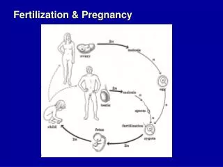
Fertilization & Pregnancy • Fertilization and Implantation
Early development of Zygote • Fertilization occurs in the oviduct
IB LEARNING OBJECTIVE • Outline early embryo development up to the implantation of the blastocyst.
Early development of Zygote • After fertilization, the zygote undergoes cleavage and develops into a blastocyst before implantation in the endometrium
Early development of Zygote • Zygote is transplant to the uterus by ciliary (small moving hair-like proteins) action in the oviduct • The fertilized egg undergoes cleavage. • Cleavage is the mitotic division of the zygote into a mass of daughter cells.
Early development of Zygote • When the embryo reaches the uterus is has undergone both cleavage and has formed a blastocysts. • A blastocyst is a tiny solid ball of cells.
Early development of Zygote • In humans, by 7 days the blastocysts consists of about 100 cells • It becomes imbedded in the endometrium, a process called implantation.
Early development of Zygote • The inner cell mass of the blastocyst eventually becomes the fetus. • Once implanted the embryo starts to receive nutrients from the endometrium of the uterus wall.
IB LEARNING QBJECTIVE • State that the fetus is supported and protected by the amniotic sac and amniotic fluid.
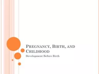
Early development of Zygote • Pregnancy, or gestation, is the condition of carrying one or more embryos in the uterus
Gestation – zygote to embryo to fetus in human • Gestation – the period of development in the mother’s body, lasting from conception to birth. • First 2 months of gestation the developing baby is referred as an embryo. • The embryo is protected in amniotic fluid and amniotic sac.
Early Development Amniotic sac Placenta • A fluid-filled amniotic sac, which cushions and protects the developing embryo. Umbilical cord Uterus Amnion Fetus
Early Development –Amniotic Fluid Amniotic sac Placenta • The fetus floats in the amniotic fluid. • This fluid acts as a shock absorber. Umbilical cord Uterus Amnion Fetus
IB LEARNING OBJECTIVE • State that materials are exchanged between the maternal and fetal blood in the placenta.
During its first 2 to 4 weeks, the embryo obtains nutrients directly from the endometrium • Meanwhile, the outer layer of the blastocyst mingles with the endometrium and eventually forms the placenta • Blood from the embryo travels to the placenta through arteries of the umbilical cord and returns via the umbilical vein
The outer layer of the embryo gives rise to the placenta& the maternal endometrium • It is a disc shaped structure that allows for an exchange of material between fetus and mother
The outer layer of the embryo gives rise to the placenta & the maternal endometrium It is a disc shaped structure that allows for an exchange of material between fetus and mother Maternal veins Maternal arteries Placenta Maternal portion of placenta Umbilical cord Chorionic villus containing fetal capillaries Fetal portion of placenta (chorion) Maternal blood pools Umbilical arteries Uterus Fetal arteriole Umbilical vein Fetal venule Umbilical cord
The Placenta • A disc shaped structure composed of maternal endometrial and fetal membrane. • Exchange in the placenta – is by diffusion and active transport, and involve: • Respiratory gases – Oxygen and carbon dioxide • Water • Excretory products (urea) • Antibodies ( immunity from diseases)
Maternal, foetal exchanges across placenta The foetus develops and grows using materials obtained by exchange across the placental wall from mother to child. Excretory products are exchanged in the opposite direction from child to mother.
IB Learning Objective • Explain how the structure and functions of the placenta, including its hormonal role in secretion of estrogen and progesterone, maintain pregnancy.
Structure and function of placenta • The female blood supply which supplies the foetus with oxygen and nutrient. • It will also remove waste from the foetal blood and excrete this through the maternal systems.
Structure and function of placenta a. Umbilical cord connects the fetus to the placenta b. There are two umbilical arteries that carry the deoxygenated fetal blood to the placenta.
Structure and function of placenta c. The single umbilical vein returns the oxygenated blood to the rest of the foetal circulation. d. The placenta is normally about 190mm wide and 20 mm deep. The human placenta is more deeply integrated into the maternal tissue than any other animal.
Structure and function of placenta e) The myometrium is composed on smooth muscle in the uterusthat produces the contraction in labor. f. The endometrium which is maintained through out pregnancy by progesterone. Initially from the corpus luteum and later from the placenta itself.
Structure and function of placenta g) The female blood supply which supplies the foetus with oxygen and nutrient. It will also remove waste from the foetal blood and excrete this through the maternal systems. h) Open ended blood arterioles and capillaries that produce the inter-villous 'blood lakes’. i) Inter-villus spaces filled with maternal blood. These surround the placental villi and allow for very efficient exchange.
Structure and function of placenta • Open ended blood arterioles and capillaries that produce the inter-villous 'blood lakes'. • .
Structure and function of placenta • Inter-villus spaces filled with maternal blood. These surround the placental villi and allow for very efficient exchange.
Structure and function of placenta • g) Placental-Villi with large surface area for the exchange of nutrient and waste.
Structure and function of placenta • Placental-Villi with large surface area for the exchange of nutrient and waste.
Structures that allow the Exchange of Material Across the Placenta