Download
1 / 58
610 likes | 717 Views
Learn about the skeletal system and its vital functions in the human body. Explore bone classification, structure, and its role in support, protection, movement, and more.

E N D
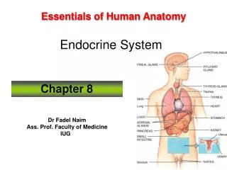
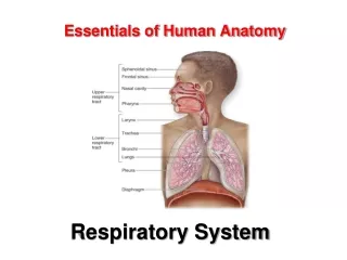
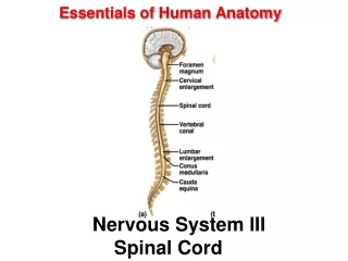
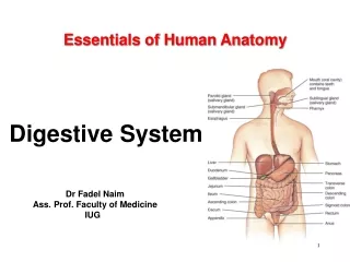
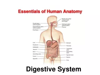
Essentials of Human AnatomyThe Skeletal System 1 Dr Fadel Naim Ass. Prof. Faculty of Medicine IUG 1
Bone • Bones are organs • Bones are composed of all tissue types. • Their primary component is osseous connective tissue. • The matrix is sturdy and rigid due to calcification (also called mineralization).
Function of Bones • Support: form the framework that supports the body and cradles soft organs • Protection: provide a protective case for the brain, spinal cord, and vital organs • Movement: provide levers for muscles • Mineral storage: reservoir for minerals, especially calcium and phosphorus • Blood cell formation: hematopoiesis occurs within the marrow cavities of bones • Energy storage (fat in yellow marrow)
Support and Protection • Bones provide structural support and serve as a framework for the entire body. • Bones protect many delicate tissues and organs from injury and trauma.
Movement • Muscles attach to the bones of the skeleton • contract and pull on bone • functions as a series of levers.
Storage of Mineral and Energy Reserves • More than 90% of the body’s reserves of the minerals calcium and phosphate are stored and released by bone.
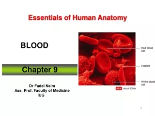
HematopoiesisBlood Cell Formation • Blood cell production in red bone marrow • located in some spongy bone. • Red bone marrow contains stem cells • form all of the blood cell types.
Bone Classification • Long Bones • Short Bones • Flat Bones • Irregular Bones • Sesamoid (Round) Bones
Classification of Bones: By Shape • Long bones – longer than they are wide (e.g., humerus)
Classification of Bones: By Shape • Short bones • Cube-shaped bones of the wrist and ankle • Bones that form within tendons (e.G., Patella)
Classification of Bones: By Shape • Flat bones – thin, flattened, and a bit curved (e.g., sternum, and most skull bones)
Classification of Bones: By Shape • Irregular bones – bones with complicated shapes (e.g., vertebrae and hip bones)
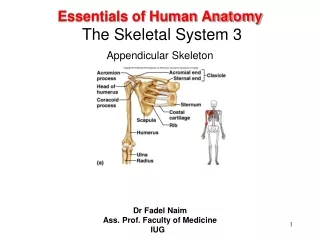
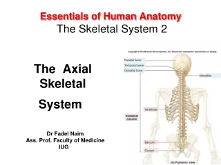
Classification of Bones • Axial skeleton – • bones of the skull, vertebral column, and rib cage • Appendicular skeleton – • bones of the upper and lower limbs, shoulder, and hip
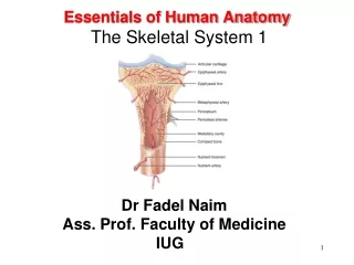
Bone Structure - External • Cartilage protection for joints
Bone Structure - External • Epiphyses • Expanded ends of long bones • Exterior is compact bone, and the interior is spongy bone • Joint surface is covered with articular (hyaline) cartilage • location of red bone marrow • Epiphyseal line separates the diaphysis from the epiphyses Epiphyse
Bone Structure - External • Diaphysis • Tubular shaft that forms the axis of long bones • Composed of compact bone that surrounds the medullary cavity • Yellow bone marrow (fat) is contained in the medullary cavity Diaphysis
Bone Structure - Internal • Spongy Bone- red marrow
Bone Structure - Internal • Compact bone
Bone Structure - Internal • Medullary Cavity-yellow marrow
Bone Structure - Internal • Epiphiseal Plate • “Growth Plate”
Parts of a Long Bone • epiphysis • distal • proximal • diaphysis • compact bone • spongy bone • articular cartilage • periosteum • endosteum • medullary cavity • trabeculae • marrow • red • yellow
Structure of Short, Irregular, and Flat Bones • Thin plates of periosteum-covered compact bone on the outside with endosteum-covered spongy bone on the inside • Have no diaphysis or epiphyses • Contain bone marrow between the trabeculae
Bone Membranes • Periosteum – double-layered protective membrane • Outer fibrous layer • dense regular connective tissue • Inner osteogenic layer • composed of osteoblasts and osteoclasts • Richly supplied with nerve fibers, blood, and lymphatic vessels, which enter the bone via nutrient foramina • Secured to underlying bone by Sharpey’s fibers • Endosteum– delicate membrane covering internal surfaces of bone
Blood and Nerve Supply of Bone • Periosteal arteries • Supply periosteum • Nutrient arteries • Enter through nutrient foramen • Supplies compact bone of diaphysis & red marrow • Metaphyseal & epiphyseal aa • Supply red marrow & bone tissue of epiphyses
Bone Markings: Projections ( Sites of Muscle and Ligament Attachment) • Tuberosity – rounded projection • Crest – narrow, prominent ridge of bone • Trochanter – large, blunt, irregular surface • Line – narrow ridge of bone
Bone Markings: Projections ( Sites of Muscle and Ligament Attachment) • Tubercle – small rounded projection • Epicondyle – raised area above a condyle • Spine – sharp, slender projection • Process – any bony prominence
Bone Markings: Projections That Help to Form Joints • Head – bony expansion carried on a narrow neck • Facet – smooth, nearly flat articular surface • Condyle – rounded articular projection • Ramus – armlike bar of bone
Bone Markings: Depressions and Openings • Meatus – canal-like passageway • Sinus – cavity within a bone • Fossa – shallow, basinlike depression • Groove – furrow • Fissure – narrow, slitlike opening • Foramen – round or oval opening through a bone
The Histologic Types: • Compact bone ( cortical) • Spongy bone ( cancellous) • Lamellar bone: regular – mature • Woven bone: irregular – immature – fetus - fracture • Osteoid • Callus: fracture healing
Compact Bone: • Haversian system, or osteon – the structural unit of compact bone • Lamella – weight-bearing, column-like matrix tubes composed mainly of collagen • Haversian, or central canal – central channel containing blood vessels and nerves • Volkmann’s canals – channels lying at right angles to the central canal, connecting blood and nerve supply of the periosteum to that of the Haversian canal
Compact Bone • Osteocytes – mature bone cells • Lacunae – small cavities in bone that contain osteocytes • Canaliculi – hairlike canals that connect lacunae to each other and the central canal
Spongy (cancellous) Bone • Does not contain osteons. • trabeculae surrounding red marrow spaces
Cell Types of Bone 4 types of cells in bone tissue • Osteoprogenitor cells: • Undifferentiated cells • Can divide become osteoblasts • Found in inner layer of periosteum and endosteum • Osteoblasts: • Form matrix & collagen fibers but can’t divide • Osteocytes: • Mature cells that no longer secrete matrix • Osteoclasts: • Huge cells from fused monocytes (WBC) • Function in bone resorption at surfaces such as endosteum
Bone Development Osteogenesis and ossification: • The process of bone tissue formation, which leads to: • The formation of the bony skeleton in embryos • Bone growth until early adulthood • Bone thickness, remodeling, and fracture repair
Bone Growth - Ossification • Cartilage template laid down. • Osteoblasts (bone building cells) located in Ossification Centers.
Bone Growth - Ossification • Primary Ossification Center in diaphasis. • Secondary Ossification Centers in epiphisis.
Bone Growth - Ossification • Grow toward one another, cartilage remains between them. • As long as cartilage remains undamaged, growth can occur.
Formation of the Bony Skeleton • Begins at week 8 of embryo development • Intramembranous ossification – bone develops from a fibrous membrane • Endochondral ossification – bone forms by replacing hyaline cartilage
Endochondral Ossification • Begins in the second month of development • Uses hyaline cartilage “bones” as models for bone construction • Requires breakdown of hyaline cartilage prior to ossification
Stages of Endochondral Ossification • Formation of bone collar • Cavitation of the hyaline cartilage • Invasion of internal cavities by the periosteal bud, and spongy bone formation • Formation of the medullary cavity; appearance of secondary ossification centers in the epiphyses • Ossification of the epiphyses, with hyaline cartilage remaining only in the epiphyseal plates
Secondary ossification center Articular cartilage Epiphyseal blood vessel Spongy bone Deteriorating cartilage matrix Hyaline cartilage Epiphyseal plate cartilage Spongy bone formation Primary ossification center Medullary cavity Bone collar Blood vessel of periosteal bud Formation of bone collar around hyaline cartilage model. 1 Cavitation of the hyaline cartilage within the cartilage model. 2 Invasion of internal cavities by the periosteal bud and spongy bone formation. 3 Formation of the medullary cavity as ossification continues; appearance of secondary ossification centers in the epiphyses in preparation for stage 5. 4 Ossification of the epiphyses; when completed, hyaline cartilage remains only in the epiphyseal plates and articular cartilages 5 Stages of Endochondral Ossification
Long Bone Growth and Remodeling • Growth in length – cartilage continually grows and is replaced by bone • Remodeling – bone is resorbed and added by appositional growth
Bone Growth in Length • Epiphyseal plate or cartilage growth plate • Cartilage cells are produced by mitosis on epiphyseal side of plate • Cartilage cells are destroyed and replaced by bone on diaphyseal side of plate • Between ages 18 to 25, epiphyseal plates close • Cartilage cells stop dividing and bone replaces the cartilage (epiphyseal line) • Growth in length stops at age 25
Remodeling • Occurs all the time. • Stresses change, bones adapt. • Osteoclasts remove bone, Osteoblasts build bone, Osteocytes maintain bone • Mineral deposits in Spongy Bone form to hold the stress best. • 5-10% bone / year.
Control of Remodeling • Two control loops regulate bone remodeling • Hormonal mechanism maintains calcium homeostasis in the blood • Mechanical and gravitational forces acting on the skeleton
Developmental Aspects of Bones • The embryonic skeleton ossifies in a predictable timetable that allows fetal age to be easily determined from sonograms • At birth, most long bones are well ossified (except for their epiphyses) • By age 25, nearly all bones are completely ossified • In old age, bone resorption predominates
Fracture • A disruption in the integrity of a living bone involving injury to: • Bone • Bone marrow • Periosteum • Adjacent soft tissues