Download
1 / 37
950 likes | 4.2k Views
cardiovascular assessment. NURS 347 Towson University. structure and function. cardiovascular system. Blood flow through the Heart. Systole & Diastole. Diastole: ventricles relax and fill with blood; this takes up two thirds of cardiac cycle

E N D
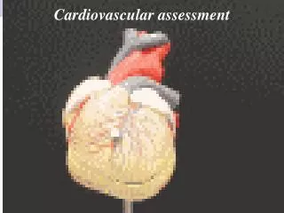
cardiovascular assessment NURS 347 Towson University
structure and function cardiovascular system
Systole & Diastole • Diastole: ventricles relax and fill with blood; this takes up two thirds of cardiac cycle • Systole: heart’s contraction, blood pumped from ventricles fills pulmonary and systemic arteries; this is one third of cardiac cycle
Systole • Ventricular pressure becomes higher than that in atria, so mitral and tricuspid valves close • Closure of AV valves contributes to first heart sound (S1) and signals beginning of systole • AV valves close to prevent any regurgitation of blood back up into atria during contraction • For a very brief moment, all four valves are closed and ventricular walls contract
Diastole • Ventricles relaxed, and AV valves, tricuspid and mitral, are open; opening of normal valve is silent • Pressure in atria higher than that in ventricles, so blood pours rapidly into ventricles • Toward end of diastole, atria contract and push last amount of blood into ventricles, known as pre-systole, or atrial systole
Cardiac Conduction • Heart has unique ability: automaticity • Can contract by itself, independent of any signals or stimulation from body • Specialized cells in sinoatrial (SA) node, near superior vena cava initiate an electrical impulse • Because SA node has intrinsic rhythm, it is called the pacemaker
PQRST • P wave: depolarization of atria • P-R interval: from beginning of P wave to beginning of • QRS complex (time necessary for atrial depolarization plus time for impulse to travel through AV node to ventricles) • QRS complex: depolarization of ventricles • T wave: repolarization of ventricles
Blood Circulation • In resting adult, heart normally pumps between 4 and 6 L of blood per minute throughout body
The Cardiovascular Assessment Peripheral Assessment
Subjective Assessment • Chest pain? • Dyspnea? • Orthopnea? • Cough? • Fatigue? • Cyanosis or pallor? • Edema? • Nocturia? • Leg pain or cramps? • Skin changes on arms or legs? • Swelling? • Lymph node enlargement? • Personal habits and self-care • Past cardiac history of self and family? • Medications
Inspection • Begin with the hands, noting: • Color of skin and nail beds • Temperature • Texture • Skin turgor • Lesions & Scars • Edema • Hair growth • Clubbing • Symmetry of extremities
Palpation • Capillary refill: • Depress and blanch the nail beds; release and note the time for color return • Color should return in less than 1-2 seconds • Pulses: Note rate, rhythm, elasticity of vessel wall, equality, and force: • 4+ Bounding • 3+ Increased • 2+ Normal • 1+ Weak • 0 Absent
Pulses • Temporal • Carotid • Radial • Ulnar • Brachial • Femoral • Popliteal • Posterior tibial • Dorsalispedal
carotid artery: palpation • Palpate each separately medial to the sternomastoid muscle of the neck • Avoid excessive pressure, may slow heart rate • Assess contour and amplitude of pulse; • Generally: • smooth contour • rapid upstroke • slower downstroke • strength 2+
carotid artery: auscultation • Middle-aged, older, or demonstrate signs and symptoms of cardiovascular disease • Listen with the bell to each side separately for a bruit • Bruit: Blowing, swishing sound Auscultation Locations: • Angle of the jaw • Midcervical area • Base of neck
Homan’s Sign • Assesses for DVT • Signs and Symptoms of DVT: • Pain & cramps • Unilateral edema • Weakened pulse • Bring leg up, allow fluid to drain. • Dorsiflex foot while squeezing the calf. • If pain is felt, positive test. • Has fallen out of favor in comparison to diagnostic imaging tests: ultrasound
cardiac exam Straight to the Heart
Inspection: precordium • Inspect for lifts, heaves, or visual pulsations: • Apical impulse • Continue to assess the integument
palpation • Palpate for lifts, heaves, thrills, or pulsations • Apical pulse (point of maximal impulse or PMI) • Use one fingerpad • 4th or 5th intercostal space, midclavicular line • Normally 1cm x 2cm and feels like a short, gentle tap • Duration is short, generally occupies only the first half of systole
palpation • General appraisal of precordium for additional pulsations: • Use the carotid artery pulsation as a guide • Use palmar aspect of four fingers and palpate: • Apex • Left sternal border • Base
auscultate • Rate & rhythm • Identify S1 & S2 • Assess each separately • Listen for extra heart sounds • Listen for murmurs • Diaphragm: High pitched sounds’ • Bell: Low pitched sounds • Use a “Z-pattern” from base of the heart, right and left, and over the apex.
Aortic: 2nd right interspace • Pulmonic: 2nd left interspace • Tricuspid: Left lower sternal border • Mitral: 5th interspace near midclavicular line • Erb’s point: 3rd interspace, left sternal border
auscultate • Heart Rate: 60-100 bpm • Rhythm: Regular (normal sinus rhythm) • Arrhythmia: varies with person’s breathing, increasing at the peak of inspiration and slowing with expiration • Pulse deficit: Auscultating apical beat and simultaneously palpating radial pulse
heart sounds: “lub-dub” • S1: “lub” • Start of systole, caused by the close of AV valves • Louder at apex • Use diaphragm • Carotid artery pulse • Electrical conduction: R- wave • S2: “dub” • Closure of the semilunar valves • Louder at the base • Can auscultate with diaphragm
extra heart sounds • Listen with the diaphragm, then the bell • Cover all auscultatory areas • Generally silent • Most common extra sound is a mid-systolic click in systole • S3: • During diastole • Ventricular gallop • S4: • During diastole • Atrial gallop
murmurs • Murmur: A blowing, swooshing sound related to turbulent blood flow in the heart or great vessels • Aside from that “innocent” murmur; murmurs are abnormal Document findings by: • Timing: During what part of the cardiac cycle? • Early, mid-, or late systole or diastole? • Throughout cardiac cycle • Muffles heart sounds?
murmur grading • Loudness: Describe intensity with the following scale: • Grade i: Barely audible, heard only in a quiet room and then with difficulty • Grade ii: Clearly audible, but faint • Grade iii: Moderately loud, easy to hear • Grade iv: Loud, associated with a thrill palpable on the chest wall • Grade v: Very loud, heard with one corner of the stethoscope lifted off the chest wall • Grade vi: Loudest, still heard with entire stethoscope lifted just off the chest wall
murmurs • Pitch:Describe as “high, medium, or low” • Depends on the pressure and rate of blood flow producing the murmur • Pattern: Does it follow a pattern through the cardiac phase? • Crescendo: Grows louder • Decrescendo: Tapers off • Crescendo-decrescendo: Increases to a peak and then decreases • Quality: • Musical • Blowing • Harsh • Rumbling • Location: • Describe the area where the murmur is best heard: • Valve area • Intercostal spaces
murmurs • Radiation: May be transmitted in the direction of blood-flow: • Another precordial area • Neck • Back • Axilla • Posture:Some murmurs disappear or are enhanced by a change in position. • Innocent: No valvular or other pathologic cause • Generally soft, grade ii, midsystolic, short • Crescendo-decrescendo, musical quality • 2nd or 3rd intercostal space, disappears with sitting • No history of cardiac dysfunction • Functional: Due to increased blood flow of the heart
physiologic splitting • A split S1 means you are hearing the mitral and tricuspid components separately, it is “normal” • Very rapid; 0.03 seconds apart • Auscultate over the tricuspid valve area • A split S2 occurs toward the end of inspiration, is normal. • “T-DUB” • Auscultate over the pulmonic valve area, 2nd interspace • Fixed split: Unaffected by respiration, always there • Paradoxical split: Sounds fuse on inspiration; split on expiration
Jugular Venous Distention (JVD) • A technique used to assess central venous pressure (CVP) • Judges the heart’s efficiency as a pump • Position patient supine at a 30-45’ angle • Remove pillow to decrease flexing the neck • Turn head slightly from area being assessed • Look for pulsating internal jugular veins near the suprasternal notch, or origin of the sternomastoid muscle near the clavicle • Be careful not to confuse the carotid pulse with the internal jugular
variations • Infants • Fetal shunt closure may take up to 48 hours • Apical pulse may be palpable at 4th intercostal space, lateral to midclavicular line. • HR 100-180 bmp after birth; 120-140 bmp average • Sinus arrhythmias with respirations • Children • Apical pulse palpation changes • 70-100 bmp • Venous hum • Innocent murmurs
variations • Pregnant female: • Increased resting heart rate • Mild hyperemia (increased cutaneous blood flow to eliminate excess heat) • Increased volume of S1, exaggerated S1 split • Heart murmurs • Aging Adult: • Gradual rise in blood pressure • Orthostatic hypotension • Avoid pressure on carotid artery • Decreased visibility of JVD • Ectopic beats
sample charting • Subjective: • No chest pain, dyspnea, orthopnea, cough, or edema. No past history of hypertension, abnormal blood tests, heart murmur, or rheumatic fever in self. Last ECG 2 years. PTA, result normal. No stress ECG or other heart tests. • Objective: • Neck: Carotids 2+ and = bilaterally, internal jugular vein pulsations present when supine, and disappear when elevated at a 45’ position. • Precordium: Inspection. No visible pulsations, no heave or lift. • Palpation: Apical pulse in 5thicsat left midclavicular line, no thrill • Auscultation: Rate 68 beats per minute, rhythm regular. S1-S2 present, not diminished or accentuated, no S3, no S4, no other extra heart sounds, no murmurs.