Comprehensive Methods for Quantitative Flotation and Larvae Harvesting in Parasitology
130 likes | 243 Views
This document outlines advanced techniques for the quantitative flotation of eggs and the harvesting of L3 larvae from fecal samples in parasitology. It includes detailed methodologies like using flotation liquids, centrifugation, and visual examination of eggs per gram (EPG) for accurate parasite detection. The Baermann technique is explained for isolating lungworm larvae, alongside methods for identifying specific parasites such as Nematodirus and Cryptosporidium spp. These processes are vital for effective diagnosis and treatment in veterinary parasitology.

Comprehensive Methods for Quantitative Flotation and Larvae Harvesting in Parasitology
E N D
Presentation Transcript
Quantitative Flotation (McMaster)
56 ml Gauze 30x30 cm Stir Wait 30 min. 4 gram Remove supernatant with pipette Fill a reading chamber Wait 5 min. Read 3 vertical lines at x200 magnification and note species and Count numbers Multiplication factor: 47 Add 3ml flotation liquid and mix with pipette 10 ml 3 ml Centrifuge 1200 g 5-7 min
x20 Added flotation liquid 1,5 / 3 / 6 ml 3 ml Visual area EPG (Eggs Per Gram) = Count X Multiplication factor example. EPG = 2 x 83= 166
40 U = 8
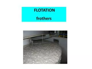
Sedimentation (heavy parasite eggs)
Sedimentation („Trematode“ eggs) Method for heavier eggs that do not flotate in common flotation fluids Nematodirus spp. Fasciola hepatica Dicrocoelium dendriticum Fasciola buski Opisthrchis noverca 3 g faeces + 50 ml water Fill with water Wait 10 min Remove supernatant gauze Add drop to slide + cover glass + read Add 1 drop of dye + mix Remove supernatant Wait 10 min Resuspend in water
Baermann (lungworms & L3)
Baermann (lungworms & L3 larvae) Harvesting of larvae from faecal sample Fold gauze and put in syringe Fill with water Wait 24 hours (no direct sun) 5-10 g faeces water Remove faeces and excess water Transfer to 14 ml centrifuge tube Centrifuge 1000 rpm, 2 min Transfer to petri dish and examine under microscope Remove excess water
Baermann (lungworms) Harvesting of larvae from faecal sample Muellerius capillaris (300-320 µm) Posterior end of body often curled Dictyocaulus viviparus (390-450 µm) Cattle Dictyocaulus filaria (550-585 µm) Larvae often curved Sluggish movement Sheep Cystocaulus (340-480 µm) Additional dorsal and ventral spines on middle part of tail Protostrongylus (320-400 µm) Posterior half of body may be curled
Baermann (L3 larvae) Harvesting of larvae from faecal sample Trichostrongylus (560-796 µm) 16 gut cells, Head tapered, Sheath forms short cone Cooperia (666-956 µm) 16 gut cells, Head square with refractile bodies, Sheath tail medium tapering (Cooperia oncophora), or finely pointed and refractile (Cooperia curticei type) Ovine Bovine Ostertagia Oesophagostomum (726-923 µm) / Chabertia (650-789 µm) Approx. 32 gut cells, Head broad and rounded, Sheath tail filamentous, Larval tail not notched or lobed Strongyloides (650-850 µm) Oesophagus extends nearly half length of body, Sheath absent, Larval tail notched http://www.rvc.ac.uk/review/parasitology/RuminantL3/Introduction.htm
Ziehl-Neelsen [acid fast staining] (Cryptosporidium spp.)
Zeihl-Neelsen (Cryptosporidium spp.) Detection of C. spp. oocysts in faeces Add small amount of feaces to slide Smear feaces using second slide Dry Fixate methanol (+2% HCl) 5 min Color carbolic fuxine 20 min Add name with pencil Repeat 2 times Wash Water 10 sec Color Brilliant Green 2.5% 5 min Wash Water 10 sec Remove color 10% H2SO4 10 sec Dry Read with x400 magnification 0 = no oocysts + = 1-5 oocysts ++ = 6-25 oocysts +++ = >25 oocysts
Ziehl-Neelsen (Cryptosporidium spp.) Detection of C. spp. oocysts in faeces Read with x400 magnification 0 = no oocysts + = 1-5 oocysts ++ = 6-25 oocysts +++ = >25 oocysts http://courses.cit.cornell.edu/biomi290/microscopycases/crypto/index.htm