
The Muscular System
Note: Prefixes myo , mys , and sarco refer to muscle. The Muscular System. Naming Skeletal Muscles. Direction of muscle fibers Rectus : The muscles fascicles are parallel to the long axis of the body or limb. Transverse: The muscles fascicles are perpendicular to the long axis of

The Muscular System
E N D
Presentation Transcript
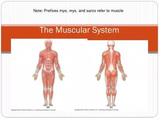
Note: Prefixes myo, mys, and sarco refer to muscle The Muscular System
Naming Skeletal Muscles Direction of muscle fibers • Rectus: The muscles fascicles are parallel to the long axis of the body or limb. • Transverse: The muscles fascicles are perpendicular to the long axis of • the body or limb. • Oblique: The muscles fascicles are aligned at an angle to the long axis of the body or limb. Location Location of a muscle Size • Maximus: Large muscle • Minimus: Small muscle • Longus: Long muscle • Brevis: Short muscle Number of origins: the end of the muscle that does not move. • Biceps: Two origins • Triceps: Three origins • Quadriceps: Four origins Shape (ie. Deltoid is triangular) Origin (proximal less movable end) & insertion (Insertion: the end of the muscle that moves) Action (What a muscle does when it contracts)
Shapes of skeletal muscles: Parallel or fusiform: • fibers run parallel to each other • contract over a great distance • good endurance but are not very strong. • Examples: Sartorius muscle and rectus abdominus muscle. Convergent: • muscle fibers converge on the insertion to maximize the force of muscle contraction • Examples: Deltoid muscle and Pectoralis Major muscle. Pennate: • many fibers per unit area. • strong but they tire quickly. • There are three types of pennate muscle. unipennate bipennate multipennete Circular: • Muscle fibers surround an opening to act as a sphincter. • Examples: Orbicularisoris and Orbicularisoculi muscles.
Do Now: • Grab your Clicker! • Review your charts and diagrams for names and locations of skeletal muscles
Id the Masseter • 1 • 2 • 3 • 4 • 5 • 6 • 7 • 8 • 9
Id the buccinator • 1 • 2 • 3 • 4 • 5 • 6 • 7 • 8 • 9
Id the orbicularisoris • 1 • 2 • 3 • 4 • 5 • 6 • 7 • 8 • 9
Id the frontalis • 1 • 2 • 3 • 4 • 5 • 6 • 7 • 8 • 9
Id the deltoid • 1 • 2 • 3 • 4 • 5 • 6 • 7 • 8. • 9
Id the sternocleidomastoid • 1 • 2 • 3 • 4 • 5 • 6 • 7 • 8. • 9
Id the external obliques • 1 • 2 • 3 • 4 • 5 • 6 • 7 • 8. • 9
Id the serratus anterior • 1 • 2 • 3 • 4 • 5 • 6 • 7 • 8. • 9
Id the rectus abdominis • 1 • 2 • 3 • 4 • 5 • 6 • 7 • 8. • 9
Id the gastrocnemius • 1 • 2 • 3 • 4 • 5 • 6 • 7
Id the biceps femoris • 1 • 2 • 3 • 4 • 5 • 6 • 7
Id the semimebranosis • 1 • 2 • 3 • 4 • 5 • 6 • 7
Id the soleus • 1 • 2 • 3 • 4 • 5 • 6 • 7
Id the semitendonosis • 1 • 2 • 3 • 4 • 5 • 6 • 7
ID the trapezius • 1 • 2 • 3 • 4 • 5 • 6
ID the infraspinatus • 1 • 2 • 3 • 4 • 5 • 6
ID the lattisimusdorsi • 1 • 2 • 3 • 4 • 5 • 6
ID the teres minor • 1 • 2 • 3 • 4 • 5 • 6
ID the brachioradialis • 1 • 2 • 3 • 4 • 5 • 6 • 7 • 8 • 9 • 0
Id the sartorius muscle • 1 • 2 • 3 • 4 • 5 • 6 • 7
Id the vastuslateralis • 1 • 2 • 3 • 4 • 5 • 6 • 7
Id the rectus femoris • 1 • 2 • 3 • 4 • 5 • 6 • 7
Why do you think that muscle-enhancing supplements (steroids, creatine) are so popular at all levels of fitness and athletics (high school, college, pro, etc.) and do you see any problems with this?
Muscle Tissues • Cardiac • Involuntary striated muscle • Dinucleated • Communicate via intercalated disks • Natural contraction cycle determined by pacemaker cells • Smooth • Lines blood vessels, digestive organs, urinary system, and parts of respiratory system • Involuntary nonstriated muscle • Ca+ triggers contractions differently (lacks sarcomeres) • Skeletal • Voluntary striated muscle • Multinucleated cells called muscle fibers • Controlled by motor nerve cells • 40% of your body mass!
Functions of Skeletal Muscle • Excitable • Receive and respond to stimulus • Contractible – Produce Movement • Pull on tendons and move bones • Maintain posture and body position • Continuous contractions maintain posture • Stabilize & strengthen joints • Support/protect soft tissues • Abdominal wall • Floor of pelvic cavity • Guard entrances and exits • Voluntary control of swallowing, defecation, and urination • Maintain body temp • Some energy from contractions lost as heat • Extensibility • Can stretch beyond its resting length • Elasticity • Recoils to resume resting length
Muscle Attachments • Most muscles attach to bones in 2 places • Insertion- movable bone • Origin – immovable/less movable bone • Direct Attachments • Epimysium of muscle fused to periosteum of bone • Indirect Attachments (more common) • Tendons – connect skeletal muscle to periosteum of bones • Aponeurosis – layers of tendons that form a sheet
Organization of Skeletal Muscle: Gross Anatomy • Each elongated cell is called a “muscle fiber” • Contains several tissues • Connective Tissue Sheaths • Epimysium – dense irregular connective tissue surrounding entire muscle • Perimysium– divide skeletal muscles into bundles of fibers (fascicles) • Endomysium – areolar tissue that surrounds ea/fiber • Blood vessels • 1 artery & 1+ veins • Numerous cross-linked capillaries • Nerves • Typically 1 nerve ending per muscle fiber • Skeletal muscle • See next page for details….
Connective tissue coverings of a skeletal muscle listed from superficial to deep are • Endomysium, perimysium, epimysium • Endomysium, epimysium, perimysium, • Epimysium, Endomysium, perimysium, • Epimysium, perimysium, Endomysium,
Microanatomy of Skeletal Muscle • Sarcolemma – plasma membrane • T tubules – transverse invaginations • Sarcoplasm – cytoplasm • Increased myoglobin • Increased gylcosomes • Myofibrils– bundles of myofilamentsparallel to cell • Thin filaments – actinproteins • Thick filaments – myosin proteins • Used for contraction • Sarcoplasmic reticulum – smooth ER (stores Ca+) • Sarcomeres– repeating units of myofilaments
Sarcomere: functional contractile unit of a muscle fiber • Sarcomere- 2µm region of myofibril between two Z-discs (Z-lines) • Striations - repeating units of dark A bands and light I bands • H zone – light stripe in the middle of the A band, with dark vertical M line • Z disc or Z line in the I band • All banding patterns are due to myofilaments: • Actin – thin filament (blue) • Myosin – thick filament (tan)
Actin (thin filaments) & Myosin (thick) • Myosin – globular heads that face outwards to cross-bridge (link) to actin, and contain ATPase • Myosin tails form central part of molecule • Cross bridges – act as motors to generate tension • Regulatory proteins • Tropomyosin – spirals around actin to block myosin binding sites in relaxed muscle • Troponin – inhibitory… helps position tropomyosin on actin and binds to Calcium ions
Sarcoplasmic Reticulum & T Tubules • Sarcoplasmic Reticulum (SR) – elaborate smooth ER like a sleeve along myofibril w/ perpendicular cross channels called terminal cisternae • T Tubules – protrude deep into cell encircling each sarcomere
Sliding Filament Theory • ACh (Acetylcholine) – neurotransmitter that released at neuromuscular junction (1 per muscle fiber) • SR & T-tubules release Ca+2into sarcoplasm • Ca+2 binds to troponin, removing blocking action of tropomyosin • Myosin heads attach to actin (cross bridge is formed) • Power Stroke: myosin pulling actin towards midline of sarcomere powered by hydrolysis of ATP ADP + Pi • Acetylcholinesterase – enzyme that breaks down Ach to prevent continued contraction • Ca+2 recaptured by SR, tropomysoin blocks binding site • Myosin heads release actin relaxing sarcoemre
The command to contract is distributed deep into the muscle fiber by • Sarcolemma • Sarcomere • Transverse tubules • myofibrils
Homework: • Compare Pallor mortis, Algor mortis,, Rigor mortis, Liver mortis, and putrification • Describe how this relates to the muscular system • Cite your source (informal URL is fine) • -OR- • Draw a diagram depicting stages of muscle contraction and relaxation labeling structural components
Energetics of Muscle Activity • Active Skeletal Muscle fiber requires 600 trillion ATP/sec! • Stored ATP- lasts 4-6 seconds • Quickly regenerated by: • Creatine Phosphate (CP) • ADP + Creatine-P ATP + creatine • Lasts 15 sec (replenishes during inactivity) • Aerobic Metabolism (Glycolysis Krebs Oxidative Phosphorylation) • Provides 30% of ATP needed during peak exertion, 95% of ATP during prolonged exertion • Glycogen, blood glucose, after 30 mins fatty acids • High yield ATP (38 ATP), but slow • Anaerobic Metabolism (glycolysis) • Main E source • Lactic acid builds up (gone 30 mins after exercise stops) • Faster but ineffective… only 2ATP • Muscle Fatigue – physiological inability to contract despite stimulation • lactic acid build up • Ioinc imbalances • Recovery Period – returns to pre-exertion levels
Please make your selection... • Choice One • Choice Two
EPOC: Excess Postexcercise Oxygen Consumption • Oxygen debt • extra O2 needed by body to restore all nonaerobic sources of ATP • Replenish O2 reserves in myoglobin • Liver must convert lactic acid to pyruvic acid • Glycogen stores must be replaced • ATP and creatine-P reserves must be resynthesized • Only 25-40% of energy released is used for work, remainder is released as heat
Muscle Mechanics • Motor unit - # muscle fibers controlled by singe motor neuron • Fine control of movement determined by size of motor unit • Ex. Eye vs. Leg muscles • During sustained contraction – motor units activated on a rotating basis (some rest/some contract) • Muscle tone -slightly contracted relaxed muscles to maintain posture and stabilize joints • Muscle twitch –response of motor unit to a single motor neuron (in lab) • Each twitch has 3 phases: • Latent period (2msec) – action pot. & release of Ca+2 • Contraction phase (peak) (10-100 msec) – cross bridges active • Relaxation phase(10-100 msec) –reuptake of Ca+2 sarcomere returns to original length
Types of Contractions • Isotonic Contractions • Concentric – muscle shortens and does work • Eccentric – muscle generates force as it lengthens • Isometric Contractions • Tension builds but muscle neither shortens or lengthens • Maintains posture
Muscle Tone • Tone – resting tension (slightly contracted) • Spinal reflexes alternate contractions of motor units • Stabilizes the position of your joints and maintains posture • Any skeletal muscle not stimulated on a regular basis will atrophy – fibers become smaller and weaker • Decreases 5% per day of inactivity! • Initially atrophy is reversible • Extreme atrophy is permanent • Paralyzed muscle may be reduced by 25% of its initial size
Muscle Performance • Force and endurance depends on: • Types of muscle fibers (most muscles contain a mixture of both but genetic variation) • Fast Twitch (white) • Powerful rapid contractions • Glycogen used quickly • Fatigue rapidly (few mitochondria) • Anaerobic, few mitochondria • Little myoglobin • Lactic acid builds up quickly • Slow Twitch (red) • Slow contraction • Aerobic, high endurance • Many mitochondria • Extensive capillary supply • Myoglobin - binds O2 reserves in muscle cell
Exercise • Anaerobic (Resistance) • Frequent, brief intense workouts • Weightlifting, isometric exercise • Results in • Hypertrophy of muscle fibers • More mitochondria • More myofilaments and myofibrils • Store more glycogen • Aerobic (Endurance) • Sustained low levels of activity • Walking, swimming, biking, jogging • Results in: • Increases # capillaries • Increases # mitochondria • Increases myoglobin synthesis • Carb-load the day before; drink glucose rich sports drinks
An activity that would require anaerobic endurance is • 50-meter dash • Pole vault • Weight lifting competition • All of the above


