Download
1 / 37
390 likes | 418 Views
Learn about diagnosis, indications, and diverse treatment modalities for giant hepatic haemangioma, a common liver tumor. Discover essential imaging techniques and potential complications associated with this benign tumor.

E N D
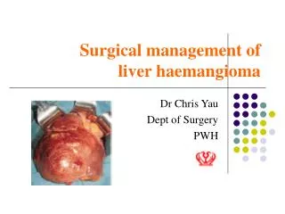
Management of Giant Hepatic Haemangioma By Dr. Hung Shun Tak Department of Surgery Princess Margaret Hospital
Introduction • First described by Ambroise Pare in 1570 • Most common benign tumors of liver • Incidence in autopsy ranging from 0.4% to 7.3%* • Age: third & sixth decade, with predominance in the fourth decades • Women to men ratio: 4:1 to 6:1** • Giant haemangioma** is defined as haemangioma with size > 4cm*** *Ishak KG, Rabin L. Benign tumors of the liver.Med Clin North Am 1975;59:995–1013. **Adam YG, Huvos AG, Forrter JG. Giant hemangiomas of the liver. Ann. Surg. 1970;172:239–245 ***Kawarada Y, Mizumoto R. Surgical treatment of giant hemangioma of the liver. Am J Surg 1984; 148:287–91
Management of Giant Hepatic Haemangioma • Presentation • Imaging • Indications • Treatment Modalities
Presentation • Asymtpomatic; uncomplicated • Pain: unclear mechanism, ? Increasing size or intratumoral thrombosis or haemorrhage secondary distension of liver capsule • Pressure symptoms: nauseas, vomiting, early satiety, or weight loss
Presentation Complication: • Spontaneous or traumatic rupture • 33 case reports of spontaneous rupture in adult, but the mortality rate is about 75%* • Kasabach Merritt Syndrome • Malignant transformation has not been reported *Ribeiro AF M., et al. Spontaneous rupture of hepatic hemangiomas: A review of the literature. World Journal of Hepatology 2010 December 27; 2(12)
Management of Giant Hepatic Haemangioma • Presentation • Imaging • Indications • Treatment Modalities
Diagnosis • Incidental findings of abdominal imagings • Majority of haemangioma would be managed non-operatively due to its benign course • Accurate diagnosis of haemangioma is essential
Imaging Source: Google picture
Ultrasonography • Heterogenous area interspersed within an hyperechoic mass Source: Márcio Martins MachadoI; Ana Cláudia Ferreira Rosa. Liver hemangiomas: ultrasound and clinical features. Radiol Bras vol.39 no.6 São Paulo Nov./Dec. 2006
Contrast Computed Tomography Source: Dario Ariel TiferesI; Giuseppe D'Ippolito. Liver neoplasms: imaging characterization. Radiol Bras vol.41 no.2 São Paulo Mar./Apr. 2008
Magnetic Resonance Imaging Source: Dario Ariel TiferesI; Giuseppe D'Ippolito. Liver neoplasms: imaging characterization. Radiol Bras vol.41 no.2 São Paulo Mar./Apr. 2008
MRI : Centripetal Enhancement Post-contrast Source: Learning radiology.com
Certainty of Diagnosis • Uncertain diagnosis due to atypical features of imagings • Diagnosis of haemangioma was established by USG in 57%, by CT scan 73% of patients* • Red blood cell ( RBC ) or MRI scans regarded as the most accurate imaging tools *Yoon SS, Charny CK, Fong Y, Jarnagin WR, Schwartz LH, Blumgart LH, et al: Diagnosis, management, and outcomes of 115 patients with hepatic hemangioma. J Am Coll Surg 2003; 197: 392–402. **Book: Surgical Management of Hepatobiliary And Pancreatic Disorders, Chapter 10: Diagnosis and management of haemangiomas of liver
Percutaneous Biopsy • Indicated in suspected malignant tumor • Risks: tumor rupture, intratumoral bleeding; seeding along the tract, intraperitoneal spread
Management of Giant Hepatic Haemangioma • Presentation • Imaging • Indications • Treatment Modalities
Does size matter? • Benign natural course/ uncomplicated • Only 33 case reports of spontaneous rupture* • No malignant transformation • Follow up in 63 patients with giant haemangioma for 33 months**: no one develop new symptom or complication, 2 haemangioma increase in size; one by 1.1cm another by 3.6cm *Ribeiro AF M., et al. Spontaneous rupture of hepatic hemangiomas: A review of the literature. World Journal of Hepatology 2010 December 27; 2(12) **Yoon SS, Charny CK, Fong Y, Jarnagin WR, Schwartz LH, Blumgart LH, et al: Diagnosis, management, and outcomes of 115 patients with hepatic hemangioma. J Am Coll Surg 2003; 197: 392–402.
Indications for Intervention 1. Incapacitating symptoms 2. Complications 3. Uncertain diagnosis 4. +/- Increase in size */** 5. +/- Size>5cm with high risk for trauma*** *Rajneesh Kumar Singh, Sorabh Kapoor, Peush Sahni, and Tushar K Chattopadhyay: Giant Haemangioma of the Liver: Is Enucleation Better than Resection? Ann R Coll Surg Engl. 2007 July; 89(5): 490–493 **Yoon SS, Charny CK, Fong Y, Jarnagin WR, Schwartz LH, Blumgart LH, et al: Diagnosis, management, and outcomes of 115 patients with hepatic hemangioma. J Am Coll Surg 2003; 197: 392–402 ***Yamagata M, Kanematsu T, Matsumata T, Utsunomiya T, Ikeda Y, Sugimachi K Management of haemangioma of the liver: comparison of results between surgery and Observation. Br J Surg. 1991;78(10):1223
Incapacitating Symptoms • 54% of patients were ultimately found to have other causes for their symptoms* • Causes: peptic ulcer disease, ischemic heart disease, gallstones or acidic reflux *Yoon SS, Charny CK, Fong Y, Jarnagin WR, Schwartz LH, Blumgart LH, et al: Diagnosis, management, and outcomes of 115 patients with hepatic hemangioma. J Am Coll Surg 2003; 197: 392–402.
Complications • Spontaneous or traumatic rupture • 33 case reports of spontaneous rupture in adult, but the mortality rate is about 75%* • Kasabach Merritt Syndrome: consumptive coagulopathy • Malignant transformation has not been reported *Ribeiro AF M., et al. Spontaneous rupture of hepatic hemangiomas: A review of the literature. World Journal of Hepatology 2010 December 27; 2(12)
Uncertain Diagnosis • Imaging: atypical appearance • High risks patients: History of colorectal cancer, HBV / HCV carrier • Resection is indicated in case of suspicion
Management of Giant Hepatic Haemangioma • Presentation • Imaging • Indications • Treatment Modalities
Treatment Modalities 1. Surgery (Resection / Enucleation) 2. Radiation therapy 3. Embolization 4. Radiofrequency Ablation
1. Surgery Resection vs Enucleation
Resection vs Enucleation • Resection: Anatomaical & non-anatomatical • In 1988, Alper et al * described a new technique : Enucleation= dissect along the fibrous cleavage plane between the capsule of haemangioma and surrounding normal liver tissue • Avoid the need to resect normal liver parenchyma and minimize damage to blood vessels and bile ducts *Aydin Alper, MD; Orhan Ariogul, MD; Ali Emre, MD; Ali Uras, MD; Attila Okten, MD. Treatment of Liver Hemangiomas by Enucleation. Arch Surg. 1988;123(5):660-661.
Resection vs Enucleation Choice depends on certainty of diagnosis and anatomical considerations
Resection vs Enucleation Indications for Resection: • potentially malignant lesions • lesions that totally replace an anatomical section of liver • Deep seated lesions • Expectant difficulty to enucleate Indications for Enucleation: • indicated in anterior and peripheral haemangioma
Resection vs Enucleation *Susan M. Lerner, MD; Jonathan R. Hiatt, MD; Johanna Salamandra, RN, BSN. Giant Cavernous Liver Hemangiomas: Effect of Operative Approach on Outcome. Arch Surg. 2004;139:818-823. **Erhan Hamaloglu, Hasan Altun, Arif Ozdemir and Ahmet Ozenc. Giant Liver Hemangioma: Therapy by Enucleation or Liver Resection. World J Surg. 2005 Jul;29(7):890-3 ***Rajneesh Kumar Singh, Sorabh Kapoor, Peush Sahni, and Tushar K Chattopadhyay: Giant Haemangioma of the Liver: Is Enucleation Better than Resection? Ann R Coll Surg Engl. 2007 July; 89(5): 490–493
Resection vs Enucleation (Conclusion) • Enucleation is preferred when feasible, as it preserves hepatic parenchyma and minimizes complications. • Resection is reserved for lesions that cannot be enucleated safely.
2. Radiation Therapy • Indication: Diffuse, multiple, massive unresectable haemangioma with symptom or progression in size • Reductions in volume from 20-40%, with 30% improvement in symptomatology • Mechanism: provoke sclerosis of tumor parenchyma with subsequent reduction in size • Complication: hepatitis, centrilobular thrombosis, tumor rupture, malignant transformation*, SCC in kidney* *Michael J. McKay, Peter J. Carr2, Allan O. Langlands. Treatment of hepatic cavernous haemangioma with radiation therapy: case report and literature review.Aust N Z J Surg. 1989 Dec;59(12):965-8.
3. Embolisation Indication: - symptomatic giant haemangioma - complicated with coagulopathy - preoperative embolization * Result: reduction in tumor size**, symptomatology** correction of coagulopathy*** *Akamatsu N, Sugawara Y, Komagome M, Ishida T, Shin N, Cho N, Ozawa F, Hashimoto D. Giant liver hemangioma resected by trisectorectomy after efficient volume reduction by transcatheter arterial embolization: a case report. J Med Case Reports. 2010 Aug 23;4:283. **Giavroglou C, Economou H, Ioannidis I. Arterial embolization of giant hepatic hemangiomas. Cardiovasc Intervent Radiol. 2003 Jan-Feb;26(1):92-6. ***EPY Fung, WH Luk, TK Loke, JCS Chan. Kasaback-Merritt Syndrome Treated by Transarterial Embolisation of Giant Cavernous Haemangioma. Transarterial Embolisation of Cavernous
Complications • Post-embolisation pain, fever, leukocytosis • Serious complications like infarction , intrahepatic abscess , sepsis are uncommon
4. Radiofrequency Ablation • Case reports of single or multiple electrode RF technology. • Mechanism: thrombogenic effect by damaging the layer of endothelial lining cause thrombosis • Complications*: ~2%, include infection, bleeding, injury to blood vessels, bile ducts, diaphragm, other abdominal organ * Ronald J. Zagoria1, Todd J. Roth1, Edward A. Levine2 and Peter V. Kavanagh. Radiofrequency Ablation of a Symptomatic Hepatic Cavernous Hemangioma . AJR 2004; 182:210-212
Summary • Hepatic hemangiomas : Most common benign tumors of the liver • Mostly asymptomatic and uncomplicated • High accuracy of diagnosis by present imaging modalities • Asymptomatic or minimally symptomatic patients can be safely observed • Indications for resection of hepatic hemangiomas include severe symptoms, inability to exclude malignancy, and complications • Enucleation, when feasible, is the technique of choice for resection, and resection can be performed with minimal morbidity and rare mortality • Other treatment modalities need further studies to support its validity