Download
1 / 39
440 likes | 947 Views
Management of Hepatic Cysts. Suen PY Department of Surgery PMH 11 February 2012. C ase P resentation. Case Presentation. Ms Cheung OL F/62 PMH: HT Past Surgical Hx: nil Social Hx: lives with daughter . Case Presentation.

E N D
Management of Hepatic Cysts Suen PY Department of Surgery PMH 11 February 2012
Case Presentation • Ms Cheung OL • F/62 • PMH: HT • Past Surgical Hx: nil • Social Hx: lives with daughter
Case Presentation • C/O: abdominal distension and right upper quadrant discomfort for 5 years with increase in severity in recent 6 months • Decrease in appetite • weight loss (5 pounds in recent 1 year)
Case Presentation-Physical Exam • no jaundice, no pallor, no cervical lymphadenopathy • Abdomen: grossly distended, hepatomegaly with liver span about 25 cm, smooth edge and no shifting dullness
Case Presentation-Laboratory Results • LFT normal (TB 22 umol/L, ALP 64 U/L, ALT 17U/L, albumin 41 g/L) • CEA: 6.1 • AFP: 1.89 • HbsAg: –ve • Hb 12.1g/dL, WBC 4.4x10⁹/L
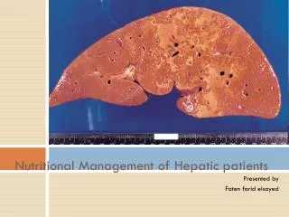
Case Presentation-Imaging • Bedside USG abdomen: huge cystic lesion in liver • USG abdomen in x-ray dept. (19/4/11): a huge liver cyst with well-circumscribed, thin and regular wall, about 24 cm in diameter over left lobe, no other liver mass
Case Presentation-imaging • CT abdomen (15/8/11): a huge liver cyst (near water density) with size of 24x15x24cm over left lobe with significant mass effect, no significant contrast enhancement in the lesion
Case Presentation • Laparoscopic liver cyst fenestration (marsupialization/unroofing) offerred; patient opted for OT • Operation done on 23/11/11 • Findings: a large left hepatic cyst (ab0ut 25 cm in diameter); about 3 litres of serous fluid inside and drained
Case Presentation-Procedure • Sub-umbilical port made under direct vision with pneumoperitoneum created; 10mm epigastric and 5mm right subcostal ports created • Cyst wall punctured and cystic fluid drained • Cyst wall partially excised • Inner lining of cyst wall cauterized • A piece of omentum anchored into cystic cavity
Case Presentation • Post-operatively: uneventful • Discharged on D5 • Followed up 1 mouth later: • Well, no more abdominal distension nor discomfort • Abdomen: soft and not distended • Wound healed • Pathology: a single layer of cuboidal epithelium, suggestive of simple hepatic cyst
Hepatic Cysts • Simple hepatic cysts (majority) • polycystic liver disease • Neoplastic cysts (benign or malignant) • Traumatic cysts • Parasitic (hydatid) cysts • Pyogenic cysts
Simple hepatic cysts-definition cystic formations of the liver, containing serous fluid, usually not communicating with biliary system
Simple hepatic cysts • Most common cystic lesions of the liver • 2nd most common incidental findings of benign lesions in the liver after hemangioma • prevalence : 5% • 90-95% asymptomatic
Simple hepatic cysts • For asymptomatic , female to male ratio about 1:1 • For symptomatic, female to male ratio 9:1 • No malignant potential • About half of patients have a single cyst, whereas the other half have two or more
Simple hepatic cysts • Pathology: Lined by a single layer of cuboidal or low columnar epithelium • Pathogenesis: regarded as a congenital malformation of aberrant bile duct, usually lost communications with biliary tree and may gradually dilate
Simple hepatic cysts-clinical presentation • Majority : asymptomatic • Commonly discovered as incidental finding during radiographic studies for unrelated symptoms or for other diseases • Common symptoms: abdominal discomfort, abdominal distension, nausea or vomiting • Rare symptoms: fever, sweating, back or shoulder pain
Simple hepatic cysts-complications • Rare • Intra-cystic haemorrhage (most common; sudden onset of increase in abdominal pain or distension ) • Spontaneous rupture • Infection • Biliary compression with obstructive jaundice • torsion
Simple hepatic cysts-diagnosis • Usually diagnosed by USG or CT • USG findings of simple hepatic cysts • Well-circumscribed • Thin and regular wall • Homogeneously anechoic • No septation, mural nodules or projections
Simple hepatic cysts-diagnosis • CT findings • Well-defined • Thin and regular wall • Homogenous, hypoattenuated fluid with density similar to water
Simple hepatic cysts-diagnosis • MRI may be considered when the diagnosis is equivocal • Well-defined, thin and regular wall • Fluid signal intensity: low on T1-weighted images and high on T2-weighted image • No wall enhancement, nodules or projections; and no internal signals
Simple hepatic cysts-diagnosis • Cyst fluid analysis ( percutaneous fluid aspiration for analysis) may also be considered in cases with difficulty in diagnosis • Cytological analysis: acellular fluid and absence of mucin • Chemical analysis: normal CEA, CA19.9 and bilirubin level
To differentiate from neoplastic cysts Neoplastic cysts’ characteristics: • Multi-locular ,septated • Thick irregular wall • Mural nodules, projections present • Thick fluid • Mucinous material in fluid • Elevated CEA or CA19.9 in fluid
Neoplastic cysts • Rare • Cystadenomas or cystadenocarcinoma • Most are cystadenomas -A benign cystic tumour with potential malignant transformation to cystadenocarcinoma (very rare) • Radiologically : complex cystic lesions
Simple hepatic cysts-treatment • Majority of patients require no treatment, just for observation
Simple hepatic cysts-indications for treatment • Symptomatic condition (most common) • Intracystic hemorrhage • Diagnostic uncertainty
Simple hepatic cysts-treatment modality • Simple percutaneous aspiration • Percutaneous aspiration followed by injection of a sclerosing agent • Fenestration (unroofing or marsupialization) • Enucleation (rarely applied)
Treatment-simple percutaneous aspiration • Percutaneous aspiration associated with very high recurrence rate (75-100%) • repeated aspiration can result in cyst infection • usually not for definitive treatment
Treatment-cyst aspiration with injection of sclerosing agent • sclerosing agents : ethanol, minocycline hydrochloride, tetracycline hydrochloride • Recurrence rate: 20-30% • contraindicated if there is communication with biliary tract • generally reserved for patients with high operative risk
Treatment-fenestration • lowest (5 %) recurrence rate • Should be considered and offered for most of symptomatic patients • A laparoscopic approach is favoured (lots of evidence demonstrates it’s treatment results equivalent to that of an open approach, while it has the advantages of a laparoscopic surgery)
Treatment-laparoscopic fenestration procedure • Laparoscopic approach adopted • Resection of a portion of the cyst wall allows drainage into the peritoneal cavity and access to its interior • Ablation of remaining inner lining of cyst wall by cauterization will minimize recurrences and the risk of ascites • A piece of omentum can be anchored into the cavity of cyst to avoid reformation of cyst
Treatment Algorithm Cystic lesion(s) Simple cystic lesion(s) complex cystic lesion(s) M.F. Hansman et al/ The American Journal of Surgery 181 (2001) 404-410 symptomatic asymptomatic polycystic liver disease simple hepatic cyst(s) Dominant cysts multiple small cysts Resect. Obs. Fenest. Fenest. Obs. Resect.
Summary (management of hepatic cysts) 1. Making a definitive diagnosis of the nature of the cystic lesion -DDx: simple hepatic cysts/neoplastic cysts/others -Inx: US/CT +/- MRI or cystic fluid analysis 2. Determining whether the patient’s symptoms are related to the cystic lesion or not -careful history taking -relevant investigations or procedures
Summary 3. Deciding whether to intervene or not -assessing the severity of symptoms, occurrence of complications, certainty of the diagnosis, pre-morbid state and the operative risks 4. Deciding the treatment modality -laparoscopic fenestration, percutaneous aspiration followed by injection of a sclerosing agent, simple aspiration or enucleation
Reference • Current Surgical Therapy by John L. Cameron, 10th ed. • Surgery of the liver and biliary tract by L.H. Blumgart, 3rd ed. • Hansman MF et al: Management and long-term follow-up of hepatic cysts, Am J Surg 181: 404-410, 2001 • Fabiani P et al: long-term outcome after laparoscopic fenestration of symptomatic simple cysts of the liver, Br J Surg 92: 596-597, 2005 • Mazza OM et al: Magagement of non-parasitic hepatic cysts, Am J Surg 209: 733-739, 2009 • www.medscape. com